 |
Case Report
Acute pancreatitis in a patient with COVID-19
1 Gastroenterology Specialist Trainee, Department of Gastroenterology, Barts Health NHS Trust, London, United Kingdom
2 Internal Medical Trainee, Department of Gastroenterology, Barts Health NHS Trust, London, United Kingdom
3 Foundation Trainee, Department of Gastroenterology, Barts Health NHS Trust, London, United Kingdom
Address correspondence to:
Saniath Akbar
50 Vicarage Road, Stratford, London, E15 4HD,
United Kingdom
Message to Corresponding Author
Article ID: 100011G01SA2020
Access full text article on other devices

Access PDF of article on other devices


How to cite this article
Akbar S, Pooni R, Pandey G. Acute pancreatitis in a patient with COVID-19. Edorium J Gastroenterol 2020;7:100011G01SA2020.ABSTRACT
Introduction: There have been reports of the novel coronavirus (COVID-19) causing non-pulmonary manifestations ranging from neurological to gastrointestinal symptoms.
Case Report: Here we present a case of a young patient who presented with epigastric pain, raised laboratory pancreatic lipase, and radiological evidence of peri-pancreatic necrosis. With history eliciting no identifiable risk factors for acute pancreatitis, the patient tested positive for COVID-19.
Conclusion: Our case highlights a possible etiological link between COVID-19 and pancreatitis.
Keywords: Coronavirus, COVID-19, Hepatobiliary, Pancreatitis
INTRODUCTION
Our understanding of the pathogenesis of the novel coronavirus is evolving at an almost exponential rate, which is important considering the increasing number of cases and deaths worldwide. With existing literature already reporting atypical presentations of COVID-19, noteworthy non-pulmonary diagnoses including liver injury, myocarditis, and stroke [1],[2],[3], these cases further highlight the potential multi-systemic nature of COVID-19.
Acute pancreatitis is characterized by inflammation of the pancreas. Several mechanisms have been proposed for its pathogenesis including increasing pancreatic duct pressure through duct obstruction (stones) and trypsin activation leading to pancreatic auto-digestion [4]. In severe acute respiratory syndrome coronavirus 2 (SARS-CoV-2) infection, it is believed the expression of the spike (S) protein and its interaction with angiotensin-converting enzyme-2 (ACE2) is implicated in the virus’s replication cycle [5]. Angiotensin-converting enzyme-2 is distributed across many tissues including the respiratory epithelium, kidneys, and the gastrointestinal (GI) tract. The presence of ACE2 protein in pancreatic cells may represent a potential link between COVID-19 and pancreatic injury [6].
CASE REPORT
A 24-year-old Romanian man with no reported medical history presented with a one-day history of sudden-onset, epigastric pain associated with nausea and vomiting. He denied any prodromal symptoms, including cough, fever, and dyspnoea, associated with COVID-19 [7]. Social history elicited the patient was a current smoker but did not consume alcohol. The patient denied taking any regular medications but mentioned of taking an over-the-counter herbal remedy containing “Silymarin,” an extract from milk thistle, with antioxidant properties [8]. There was no family history of hepatobiliary disease.
Physical examination showed normal observation parameters. Abdominal examination revealed epigastric tenderness with no clinical signs of peritonitis. Remaining clinical examination was unremarkable.
Laboratory blood tests on admission revealed a raised hematocrit of 0.55 (0.40–0.50), white cell count of 8.4 × 10⁹/L (4.0–7.0 × 10⁹/L), aminotransferase (AST) of 120 unit/L (0–40 unit/L), serum lipase of 380 unit/L (13–60 unit/L), adjusted calcium of 2.31 mmol/L (2.20–2.60 mmol/L), and C-reactive protein (CRP) of <1 mg/L (0–5 mg/L). Serum triglycerides were slightly elevated at 4.13 mmol/L (0–1.7 mmol/L). The remaining blood tests were within normal parameters. A lactate of 4.8 mmol/L (0– 1.5 mmol/L) was noted on the venous blood gas.
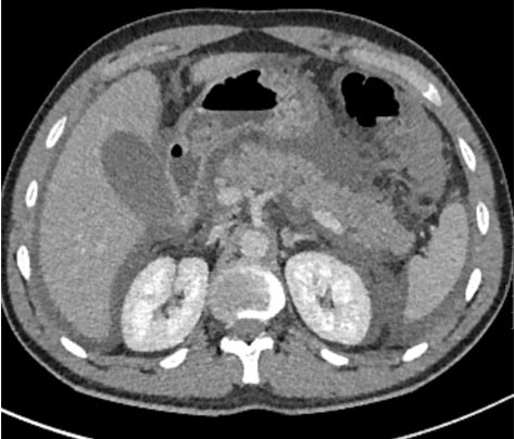
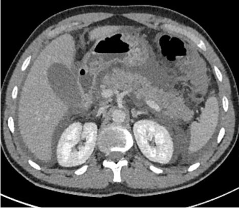
The admission chest radiograph showed no pleural effusion, pneumoperitoneum, or typical radiographic features seen with COVID-19 [9]. Although current UK guidance of acute pancreatitis discourages the use of early computerized tomography (CT) in the detection of pancreatitis [10], initial differential diagnoses including intra-abdominal pathology, namely viscus perforation, were important to rule out. Subsequently, a contrast-enhanced CT scan of the abdomen was performed showing moderate ascites with peri-pancreatic fluid, fat stranding, and areas of peri-pancreatic necrosis (Figure 1 and Figure 2).
Within 48 hours of admission, the patient’s CRP rose significantly from <1 to 372 mg/L (peaking at 410 mg/L a further 24 hours later). Within this period, the patient developed an oxygen requirement and became markedly tachycardic at a rate of 140 beats per minute. Following a prompt clinical review of the patient, polymerase chain reaction (PCR) testing for coronavirus (SARS-COV-2) was performed, which was subsequently positive.
During the admission, the patient underwent further biochemical and radiological investigations to determine the cause of his pancreatitis. Serum immunoglobulins (e.g., IgG) and alpha-1 antitrypsin levels were within normal parameters. Ultrasound of the abdomen revealed a sonographically normal gallbladder with no gallstones and a normal caliber common bile duct.
Throughout admission the patient was managed conservatively with opioid analgesia, intravenous fluids, and oxygen therapy. With tight control of blood glucose between 3.8 and 5.8 mmol/L, the patient did not require a variable rate intravenous insulin infusion (VRIII). From a nutritional point of view, the patient was managed solely with intravenous fluid hydration for the first 96 hours prior to resuming an oral diet upon discharge. There were no signs of extra-pancreatic bacterial infection, with blood cultures showing no evidence of bacteremia, and thus the patient did not receive anti-microbial therapy during admission.
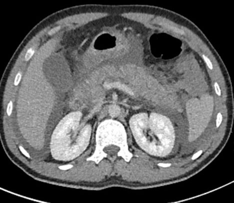
Repeat CT imaging of the abdomen prior to discharge revealed a slight increase in the moderate ascites, with peri-pancreatic fluid, fat stranding, and peri-pancreatic necrosis as before, but no evidence of parenchymal necrosis (Figure 3).
The patient did not require invasive monitoring or intensive care support and subsequently was managed on a surgical ward throughout his hospital stay. The patient made a good recovery with a total length of stay of five days.
DISCUSSION
Biliary, pancreatic, and vascular pathology are among the most common etiologies for a patient presenting with acute epigastric pain. In our case, there were concerns that a raised lactate suggested critical illness [11] and possible intra-abdominal catastrophe, prompting urgent CT imaging, which in turn showed acute pancreatitis complicated by peri-pancreatic necrosis.
Interestingly a recent case report was published of a patient with COVID-19 presenting with acute pancreatitis with no identifiable precipitating risk factors [12]. Up to 75% of cases of acute pancreatitis are due to either gallstones or alcohol [10]. In our patient case, there was no history of alcohol abuse or evidence of gallstones on ultrasonography. Upon evaluation of the patient’s marginally elevated serum triglycerides, it was deemed that they were not of clinical significance when compared to the acute pancreatic syndrome. Serum triglyceride levels are required to be above 11.2 mmol/L for a diagnosis of hypertriglyceridemia-induced acute pancreatitis to be considered [13]. Interestingly the patient was taking a Silymarin herbal of his own volition. It is commonly used by people in Southern Europe [8]. We do not think this is the cause of pancreatitis in our case. No causative link has been found between Silymarin and pancreatitis in the literature as far as we are aware. On the contrary several experimental animal studies have looked into the protective mechanism of the drug in pancreatic function and treating pancreatitis in mice [14],[15].
Although less common, an infectious etiology (viral infection) can also cause pancreatitis [10]. A drawback in our case is that we did not formally investigate viral causes, specifically mumps and Coxsackie infection. We felt there was no obvious clinical suspicion with no history of viral prodromal preceding admission. Other diagnoses to consider include autoimmune or hereditary pancreatitis, especially in such a young patient presenting with acute epigastric pain. The patient’s serum immunoglobulin G levels were normal with an absence of classic CT features one may see with autoimmune pancreatitis, making this etiology less likely [16]. With an absence of recurrent episodes of pancreatitis in his history concomitant with no history of pancreatitis in first degree relatives, a hereditary etiology is unlikely. However, with this patient being young, recurrent episodes of pancreatitis may occur in the future. In such cases, biopsy, radiographic evaluation, and genetic testing would be indicated [17],[18].
With an absence of respiratory symptoms and a normal admission chest radiograph, a diagnosis of COVID-19 was not initially considered. However, acute respiratory deterioration coupled with an increasing oxygen requirement led to nasopharyngeal swab testing for SARS-CoV-2, which was subsequently positive. Atypical presentation, in this case apparent sparing of the pulmonary system, made the diagnosis of COVID-19 a difficult one. However, it is important to remember that patients can have normal chest radiography, particularly early in the disease process; sensitivity for positive findings have been reported as low as 69% in one study [9]. Furthermore, Jin et al. [19] found that up to 28% of COVID-19 patients presenting with GI-related complaints had no respiratory symptoms.
We read with interest a retrospective analysis by Lui et al. [6] looking into the expression of ACE2 in the normal pancreas and in 121 patients diagnosed with COVID-19. The study showed high expression of ACE2 in the pancreas (e.g., the exocrine glands). In the patient cohort studied, a number of patients were found to have pancreatic injury (as defined by raised amylase/lipase and/or imaging alterations in the pancreas). The study proposed a potential mechanism of SARS-CoV-2 binding to ACE2 in the pancreas leading to pancreatic injury. This is further supported by a retrospective study from Wuhan [20] which found 17% of patients with COVID-19 pneumonia were found to have pancreatic injury (based on pancreatic enzyme levels).
It is not entirely clear if the pathogenetic mechanisms rely on viral binding to ACE2 or alternatively whether a systemic inflammatory response (SIRS) in severe COVID-19 infection maybe the cause of pancreatic injury. We believe future work needs to address the expression of SARS-CoV-2 in the pancreas of affected individuals to better delineate the association with pancreatic injury. We also believe these studies need to (diagnostically) better define patients with “pancreatitis,” fulfilling the diagnostic criteria as laid out by the UK Working Party on acute pancreatitis [10]. Indeed, as stated by de-Madaria et al. “mild increases in blood levels of pancreatic enzymes can be explained by many factors other than pancreatic damage in patients with COVID-19” [21].
CONCLUSION
In the current global coronavirus pandemic, we are seeing increasing cases of non-pulmonary manifestations of the infection. Our case highlights how a diagnosis of acute pancreatitis should increasingly be considered in our list of differential diagnoses when seeing COVID-19 patients (suspected or confirmed) presenting with epigastric pain to the Emergency Department. Could COVID-19 in the future be considered as a new etiological risk factor for pancreatitis?
ABBREVIATIONS
ACE2
AST
COVID-19
CRP
CT
GI
IgG
PCR
(S) protein
SARS-CoV-2
SIRS
VRIII
Angiotensin-converting enzyme-2
Aspartate aminotransferase
COronaVIrus Disease 2019
C-reactive protein
Computerized tomography
Gastrointestinal
Immunoglobulin G
Polymerase chain reaction
Spike protein
Severe acute respiratory syndrome coronavirus 2
Systemic inflammatory response syndrome
Variable rate intravenous insulin infusion
REFERENCE
1.
Zhang C, Shi L, Wang FS. Liver injury in COVID-19: Management and challenges. Lancet Gastroenterol Hepatol 2020;5(5):428–30. [CrossRef]
[Pubmed]

2.
Zeng JH, Liu YX, Yuan J, et al. First case of COVID-19 complicated with fulminant myocarditis: A case report and insights. Infection 2020;1–5. [CrossRef]
[Pubmed]
3.
Avula A, Nalleballe K, Narula N, et al. COVID-19 presenting as stroke. Brain Behav Immun 2020;87:115–9. [CrossRef]
[Pubmed]
4.
Steer ML. Pathogenesis of acute pancreatitis. Digestion 1997;58 Suppl 1:46–9. [CrossRef]
[Pubmed]
5.
Li Y, Zhou W, Yang L, You R. Physiological and pathological regulation of ACE2, the SARS-CoV-2 receptor. Pharmacol Res 2020;157:104833. [CrossRef]
[Pubmed]
6.
Liu F, Long X, Zou W, et al. Highly ACE2 expression in pancreas may cause pancreas damage after SARS-CoV- 2 infection. medRxiv 2020. [CrossRef]
7.
Yuki K, Fujiogi M, Koutsogiannaki S. COVID-19 pathophysiology: A review. Clin Immunol 2020;215:108427. [CrossRef]
[Pubmed]
8.
Vargas-Mendoza N, Madrigal-Santillán E, Morales-González A, et al. Hepatoprotective effect of silymarin. World J Hepatol 2014;6(3):144–9. [CrossRef]
[Pubmed]
9.
Wong HYF, Lam HYS, Fong AHT, et al. Frequency and distribution of chest radiographic findings in COVID-19 positive patients. Radiology 2019;201160. [CrossRef]
[Pubmed]
10.
Working Party of the British Society of Gastroenterology. UK guidelines for the management of acute pancreatitis. Gut 2005;54(Suppl 3):iii1–9. [CrossRef]
[Pubmed]
11.
National Institute for Healthcare and Excellence. Sepsis: Recognition, diagnosis and early management. London: NICE; 2016. (Clinical guideline [NG51]). [Available at: https://www.nice.org.uk/guidance/ng51]
12.
Anand ER, Major C, Pickering O, Nelson M. Acute pancreatitis in a COVID-19 patient. Br J Surg 2020;107(7):e182. [CrossRef]
[Pubmed]
13.
Gelrud A, Whitcomb DC. Hypertriglyceridemiainduced pancreatitis. UpToDate 2019.
14.
Kim MJ, Kim DU, Choi JW, et al. Silymarin attenuates the severity of cerulein-induced acute pancreatitis. Pancreas 2020;49(1):89–95. [CrossRef]
[Pubmed]
15.
Soto C, Mena R, Luna J, et al. Silymarin induces recovery of pancreatic function after alloxan damage in rats. Life Sci 2004;75(18):2167–80. [CrossRef]
[Pubmed]
16.
O’Reilly DA, Malde DJ, Duncan T, Rao M, Filobbos R. Review of the diagnosis, classification and management of autoimmune pancreatitis. World J Gastrointest Pathophysiol 2014;5(2):71–81. [CrossRef]
[Pubmed]
17.
Chari ST, Takahashi N, Levy MJ, et al. A diagnostic strategy to distinguish autoimmune pancreatitis from pancreatic cancer. Clin Gastroenterol Hepatol 2009;7(10):1097–103. [CrossRef]
[Pubmed]
18.
Howes N, Greenhalf W, Stocken DD, Neoptolemos JP. Cationic trypsinogen mutations and pancreatitis. Clin Lab Med 2005;25(1):39–59. [CrossRef]
[Pubmed]
19.
Jin X, Lian JS, Hu JH, et al. Epidemiological, clinical and virological characteristics of 74 cases of coronavirus-infected disease 2019 (COVID-19) with gastrointestinal symptoms. Gut 2020;69(6):1002–9. [CrossRef]
[Pubmed]
20.
Wang F, Wang H, Fan J, Zhang Y, Wang H, Zhao Q. Pancreatic injury patterns in patients with coronavirus disease 19 pneumonia. Gastroenterology 2020;S0016-5085(20):30409–1. [CrossRef]
[Pubmed]
21.
de-Madaria E, Siau K, Cárdenas-Jaén K. Increased amylase and lipase in patients with COVID-19 pneumonia: Don’t blame the pancreas just yet! Gastroenterology 2020;S0016-5085(20):30561–8. [CrossRef]
[Pubmed]
SUPPORTING INFORMATION
Acknowledgement
Dr. Kai Lee Tan, who provided the radiology input for this case report including descriptions of the images in the case.
Author ContributionsSaniath Akbar - Conception of the work, Design of the work, Drafting the work, Revising the work critically for important intellectual content, Final approval of the version to be published, Agree to be accountable for all aspects of the work in ensuring that questions related to the accuracy or integrity of any part of the work are appropriately investigated and resolved.
Rajan Pooni - Conception of the work, Design of the work, Drafting the work, Revising the work critically for important intellectual content, Final approval of the version to be published, Agree to be accountable for all aspects of the work in ensuring that questions related to the accuracy or integrity of any part of the work are appropriately investigated and resolved.
Gargi Pandey - Conception of the work, Design of the work, Drafting the work, Revising the work critically for important intellectual content, Final approval of the version to be published, Agree to be accountable for all aspects of the work in ensuring that questions related to the accuracy or integrity of any part of the work are appropriately investigated and resolved.
Guarantor of SubmissionThe corresponding author is the guarantor of submission.
Source of SupportNone
Consent StatementWritten informed consent was obtained from the patient for publication of this article.
Data AvailabilityAll relevant data are within the paper and its Supporting Information files.
Conflict of InterestAuthors declare no conflict of interest.
Copyright© 2020 Saniath Akbar et al. This article is distributed under the terms of Creative Commons Attribution License which permits unrestricted use, distribution and reproduction in any medium provided the original author(s) and original publisher are properly credited. Please see the copyright policy on the journal website for more information.


