Original Article
 
Endocytic pathway in Caco-2 cells: A model for drugs transport across the intestinal epithelial cell barrier
Mohammed A. Akeel
Chairman of the Department of Anatomy, Faculty of Medicine, Jazan University, Jazan, Kingdom of Saudi Arabia

Article ID: 100008G01MA2018
doi: 10.5348/100008G01MA2018OA

Corresponding Author:
Dr. Mohammed A. Akeel,
Faculty of Medicine, Jazan University, P.O. Box 114,
Jazan, Kingdom of Saudi Arabia

Access full text article on other devices

  Access PDF of article on other devices

[HTML Abstract]   [PDF Full Text] [Print This Article]
[Similar article in PubMed] [Similar article in Google Scholar]


How to cite this article
Akeel MA. Endocytic pathway in Caco-2 cells: A model for drugs transport across the intestinal epithelial cell barrier. Edorium J Gastroenterol 2018;5:100008G01MA2018.


ABSTRACT

Aims: Human colon adenocarcinoma (Caco-2 cells), has the ability to spontaneously differentiate towards a normal intestinal epithelium phenotype in long-term culture. Since the junctional complex, brush border, and transcytosis contribute mainly to the function of enterocytes in absorption, morphological structure of these cells plays undeniable rule. However, full morphological characterization of these cells was not adequately investigated. Therefore, the aim of the present work is to carry full morphological characterization of the cultured intestinal epithelial cells versus the Caco-2 cells model.

Methods: Freshly isolated and late cultured jejunal cells of adult rabbits as well as Caco-2 cells were processed for transmission electron microscopy (TEM) and scanning electron microscopy (SEM) and examined with Philips CM100 at 80 KV, and Philips SEM XL 30 at 30 Kv; respectively. Immune-fluorescence examination was done using two primary antibodies: anti-pancytokeratin for detecting cells of epithelial origin and anti-HBB, specific for apical uptake of sucrase-isomaltase, then processed for DAB staining. Immune-electron microscopy was done by tracing the uptake and transport of IgG conjugated to HRP, stained with DAB cytochemistry, and then processed for TEM. Results: Cultured Intestinal epithelial cells expressed early cytokeratin, tight junctional complexes, desmosomes and long microvilli with TEM before deterioration and conversion into fibroblastic like cells on day 21. Caco-2 cells in culture gradually undergo spontaneous enterocytic differentiation, in the form of cytokeratin labeling and positive sucrase-isomaltase as detected by immunofluorescence. Examination by TEM and SEM showed high density surface microvilli, coated pits and vesicles, apical tubo-vesicular system and apical junctional complexes. Immunoelectron microscopy of the Caco-2 cells showed the uptake at the surface of microvilli and transcytosis of IgG-HRP reaching the basal region.

Conclusion: This investigation provides full morphological characterization of the cultured epithelial cells and Caco-2 cells model. Caco-2 cells model has proved to be a useful in vitro model that mimics the intestinal epithelium.

Keywords: Caco-2 cells, IgG HRP, Scanning electron microscopy, Transmission electron microscopy


INTRODUCTION

Since the first attempt to culture the intestinal epithelial cells [1], it was found that many fibroblastic cells as well as epithelial cells proliferated in primary culture [2]. A key feature of the epithelial cells is the ability to maintain the specific biochemical composition of the apical and basolateral plasma membrane domains while selectively allowing transport of proteins and lipids from one pole to the opposite by transcytosis [3], [4], [5]. Clathrin-mediated endocytosis is the best characterized endocytic mechanism and is the predominant pathway for macromolecule uptake along epithelia [6], [7]. Moreover, receptor-mediated endocytosis is an essential mechanism for the transport of a variety of macromolecules into cells as well as across epithelia [8]. Fc receptors form FcRn that traffics IgG in both directions across polarized epithelial cells that line mucosal surfaces, contributing to host defense [9]. Another characteristic feature of the epithelial cells is the presence of junctional complex at the lateral cell walls, providing barrier against the penetration of noxious substances from the gut lumen [10]. These intercellular junctions associate with the peri-junctional actin cytoskeleton.

The human colon adenocarcinoma (Caco-2) cell line is an immortal culture line that spontaneously differentiates in culture to form confluent monolayers that resembles the small intestinal epithelium [11]. Caco-2 cells have been explored regarding its similarity with the intestinal epithelial characteristics [12]. Sucrase-isomaltase is an apical membrane disacharidase that is found exclusively in enterocytes of adult intestine and is expressed in a complex pattern along the intestinal crypt-villus axis [13]. The major site of expression of the enzyme sucrase-isomaltase (SI) is the brush border of the small intestine [14]. Caco-2 cells have been used extensively for analyzing the expression and biosynthesis of the enzyme sucrase-isomaltase (SI) and other brush border hydrolases [15],[16].

The Caco-2 cell line is widely used with in vitro assays to predict the absorption rate of candidate drug compounds across the intestinal epithelial cell barrier. Since the tight junctions of gastrointestinal tract epithelial cells contribute mainly to the physical barrier in absorption, morphological structure of these cells plays undeniable rule. With the widespread use of Caco-2 cell culture as a model for intestinal epithelial absorption, the question of using culture cells derived directly from the intestinal epithelium arises. However, full morphological characterization of these cells was not adequately investigated. Therefore, the aim of the present work is to investigate an in vitro long term cell culture of rabbit intestinal epithelial cells versus the Caco-2 cell line. Full morphological and functional characterization is achieved using transmission and scanning electron microscopy and immunocytochemistry. It aims also at investigating the endocytic pathway at Caco-2 cell line utilizing Goat anti-rabbit IgG conjugated to HRP.


MATERIALS AND METHODS

Isolation and culture of jejunal epithelial cells
Thirty adult New Zealand white male rabbits weighting 2–3 kg were utilized for the present study. All the animals were kept under optimum environmental and nutritional conditions. Animals were periodically assessed for general health and body weight. Ten rabbits were used for examining the normal intact morphology of the duodenal epithelium. The other twenty rabbits were equally utilized for preparing culture lines using explants isolation or collagenase isolation. At least two culture lines were prepared from each rabbit. Animals were anesthetized and the jejunum was perfused with PBS ++ under high pressure through abdominal aorta then it was taken out and emptied. The jejunum was then placed in cold (4ºC) C-199 culture media (GIBCO, Grand Island, NY) supplemented with 1% antibiotic-antimycotic (10.000 µg/ml penicillin G, 10.000 µg/ml streptomycin sulphate and 25 µg/ml amphotericin B (GIBCO, Grand Island, NY). The experimental protocol was approved by the Ethics Committee for Animal Research. Animal care was in compliance with research regulation and resolution.

Under laminar flow hood with strict antiseptic measurements, the mucosa was stripped off the underlying submucosa, and then minced into small pieces. Some of these pieces were transferred directly to culture wells (explants culture). Other pieces (1 mm3) were transferred to 1.8% collagenase and 0.05% elastase (collagenase culture) (Worthington Biochemical, Freehood, NJ). The culture medium was C-199 medium, supplemented with 1% antibiotics-antimycotic and 10% fetal bovine serum (FBS) (GIBCO, Grand Island, NY). The minced mucosa was incubated for 90 min at 37ºC in a shaking water bath. Repeated aspiration of the cell suspension was done every 15 minutes during digestion to insure cell dispersion. The cellular suspension was then filtered and pellets were washed twice in phosphate -buffered solution (pH 7.5) then resuspended in C-199 medium containing 10% FBS and 1% antibiotics -antimycotics and platted onto 25 cm2 cell culture flasks (Fisher, Springfield, NJ). They were incubated in humidified atmosphere of 95% air, 5% CO2 at 37ºC for a period of two weaks. Media were changed every other day. The cells were isolated from the floor of the culture flasks (Falcon, Heidelberg, Germany) by trypsinization. The jejunal epithelial cells were examined by the phase contrast microscope throughout all the culture periods, and the morphological structure of the crypts and cells was described.

Caco-2 cells culture
Caco-2 cells (American Type Culture Collection) (HTB-37, Manassas, VA) were obtained. Culture Collection was maintained in Dulbecco’s modified Eagle’s medium (DMEM; GIBCO, Invitrogen Ltd., Carlsbad, CA, USA) containing 2 mmol/L L-glutamine, 1 mmol/L sodium pyruvate, 10% (v/v) fetal bovine serum (FBS; GIBCO) that had been heat-inactivated for 30 minutes at 56°C, 100 U/ml penicillin, and 50 μg/ml streptomycin, and incubated at 37°C under 5% CO2-95% air atmosphere. The passages 30 to 40 were utilized in the present experiments.

Caco-2 cells were maintained at low density seeding (120–103 cells/cm2) and subcultured every six days (passage frequency). The culture medium used was Dulbecco’s modified Eagle’s minimum essential medium (DMEM) containing 25 mM glucose (Gibco Grand Island, N Y) supplemented with 20% heat-inactivated (56°C, 30 minutes) fetal bovine serum and 1% nonessential amino acids (Gibco, Glasgow, Scotland), and cultured in a 5% CO2/ 95% air atmosphere. The medium was changed every other day. Phase Contrast microscopy was used to examine the morphology of the Caco-2 cell culture throughout the various periods of culture.

Transmission electron microscopy (TEM)
The freshly isolated jejunum cells (JCs) and Caco-2 cells as well as late cultured JCs were processed for TEM study. The cells were washed twice in sterile phosphate buffer solution PBS (with Ca2+ and Mg2+, and hereafter PBS++). The cells were fixed for 60 minutes in 1% glutraldehyde, 2% paraformaldehyde in phosphate -buffer at room temperature (pH 7.4). The cells were then washed twice in buffered sucrose (0.1 M phosphate buffer, 5% sucrose solution) for five min each. Post fixation was performed at 4°C for 60 min in phosphate-buffered 2% osmium. The cells were dehydrated in a graded series of ethanol’s (40, 50, 70, 80, 90, and twice in 100%) after rinsing in several changes in cold distilled water. Embedding was carried out in Durcurpan ABCD (Sigma) in the following fashion: isolated wells were infiltrated with a 1:1 mixture of absolute ethanol and Durcurpan and allowed to stand overnight. The cells were then cured in pure Durcurpan at 35, 45 and 65°C for 12 hours at each temperature. En face and longitudinal light gold sections (1000Ä) from randomly selected series of 20 cells isolation were cut on LBK Ultratome and mounted on unsupported 300-mesh copper grids. Sections were double stained with uranyle acetate and lead citrate for 1-2 minutes and viewed at 80 KV using a Philips CM100 SEM.

Scanning electron microscopy (SEM)
The freshly isolated jejunum cells and Caco-2 cells as well as late cultured jejunal cells were utilized for EM study. The cells were washed twice in sterile PBS (with Ca2+ and Mg2+, and hereafter PBS++). The cells were fixed for 60 minutes in 1% glutraldehyde, 2% paraformaldehyde in phosphate buffer at room temperature (pH 7.4). The cells were then washed twice in buffered sucrose for five min. each (0.1 M phosphate buffer, 5% sucrose solution). Post fixation was performed at 4°C for 60 min in phosphate-buffered 2% osmium. The cells were dehydrated in a graded series of ethanol’s (40, 50, 70, 80, 90, and twice in 100%) after rinsing in several changes in cold distilled water. The cells were further dehydrated in absolute ethanol: acetone (1:1) solution for further 30 minutes, then in CPD, and coated with gold sputter coater SCD005 and then examined and photographed with Philips Scanning Electron Microscopy XL 3 at 30 Kv.

Immunofluorescence
The following primary antibodies were used at empirically derived dilution:

1- 1:200 monoclonal anti-pancytokeratin (C1801-Sigma).

2- 1:100 Monoclonal antibody anti-Hemoglobin, Beta (HBB 2/614188) (from ascites fluid) specific for sucrase-isomaltase from normal human small intestine was used (Bio Center of the University of Basel, Basel, Switzerland).

Indirect Immunofluorescence was performed on jejunal cells monolayers three days old culture and Caco-2 cells (30–40 passages) at least one month old cultures that were seeded into single well culture chamber slides (Fisher, Springfield, and N.J). The cells were fixed for 15 min at room temperature in 3.7% paraformaldehyde in Ca 2+ and Mg2+ free phosphate buffered saline.

Cells were initially incubated with blocking serum (5% goat serum 1% BSA) after washing in several changes of PBS. The culture chambers were incubated with the primary antibodies; anti–Pancytokeratin and anti-HBB in PBS, supplemented with 1% BSA for 60 minutes in a humidified 5% CO2 incubator (37°C). The secondary antibody (anti-mouse IgG FITC conjugate) was applied at 1: 128 dilution and the cells were subsequently in cubated at 37°C for 45 minutes. The cells were then washed three times in PBS at room temperature for 10 min each. The chambers were drained and disassembled and slides were mounted with Flouromount G (Fisher) and sealed with a cover slip and examined under an epifluorescence microscope equipped with a blue filter. As controls, several culture chambers were incubated with PBS and secondary (no primary) to detect non-specific binding of secondary antibody. Moreover; the cells were viewed in the absence of either primary or secondary antibodies to identify any auto-fluorescence.

Immunoelectron microscopy
The apical endocytic pathway was investigated by tracing the uptake and transport of Goat anti-rabbit IgG conjugated to horseradish peroxidase (IgG-HRP). Anti-rabbit IgG (whole molecule)- conjugated to horseradish peroxidase developed in goat (A6154) (100–200 mg/ml) was added to Caco-2 cell culture medium (2 months) at 4°C for 15 minutes after washing three Times with PBS. The Caco –2 cells were then fixed in 2% paraformaldehyde in phosphate buffer at room temperature (pH 7.4), then processed for diaminobenzidine (DAB) cytochemistry. Peroxidase reaction was initiated by incubation of Caco-2 cells in 50 mM phosphate buffer containing 0.01% H2O2 and 0.05% DAB for 10 minutes. The culture dishes were washed in PBS buffer, fixed and then processed for electron microscopy as described previously.

RESULTS

Phase contrast microscopy
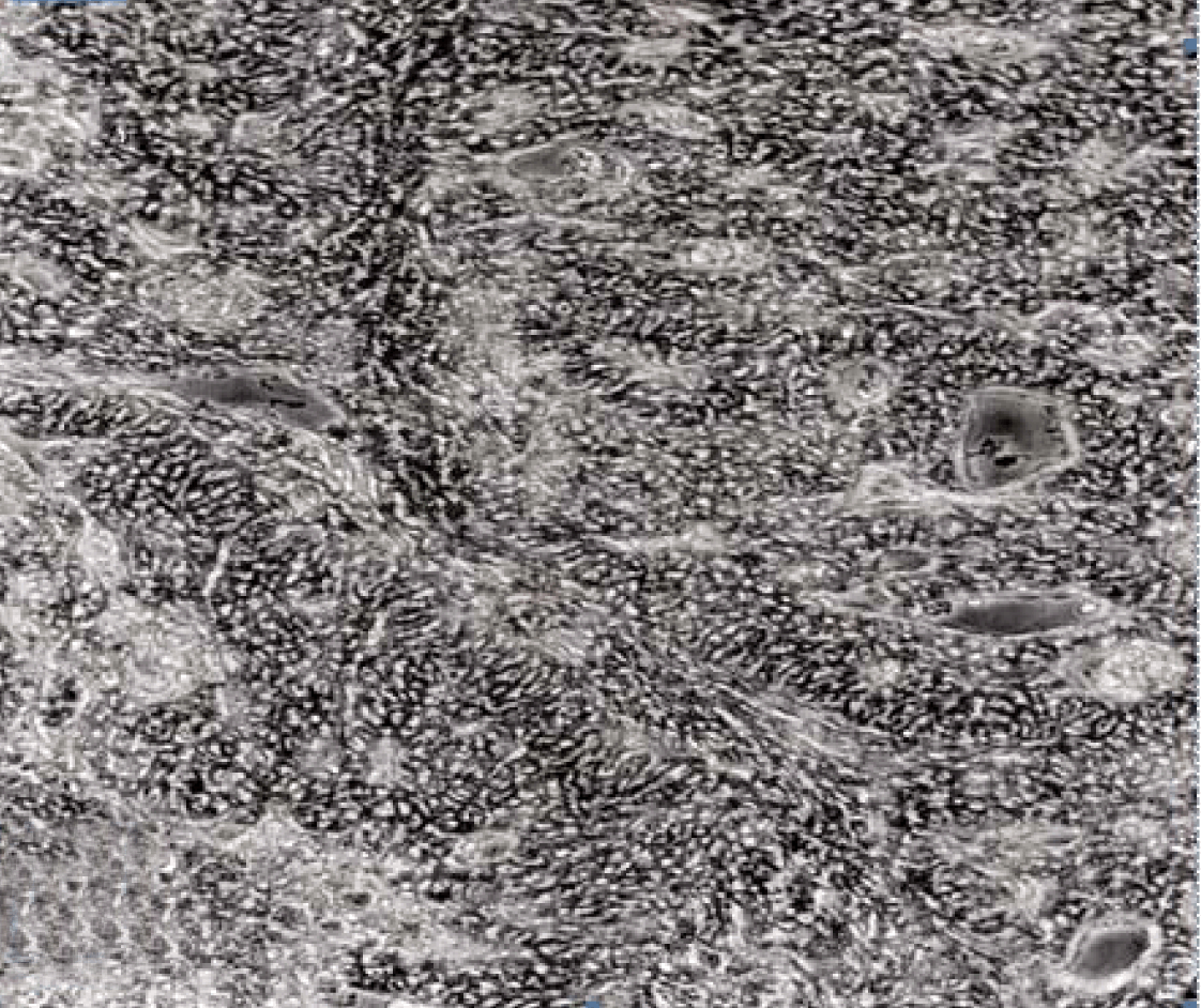
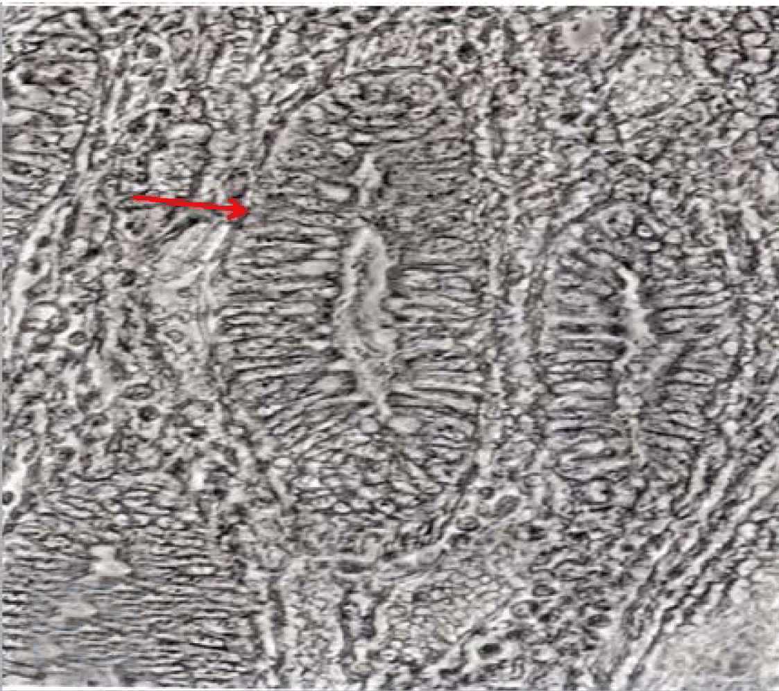
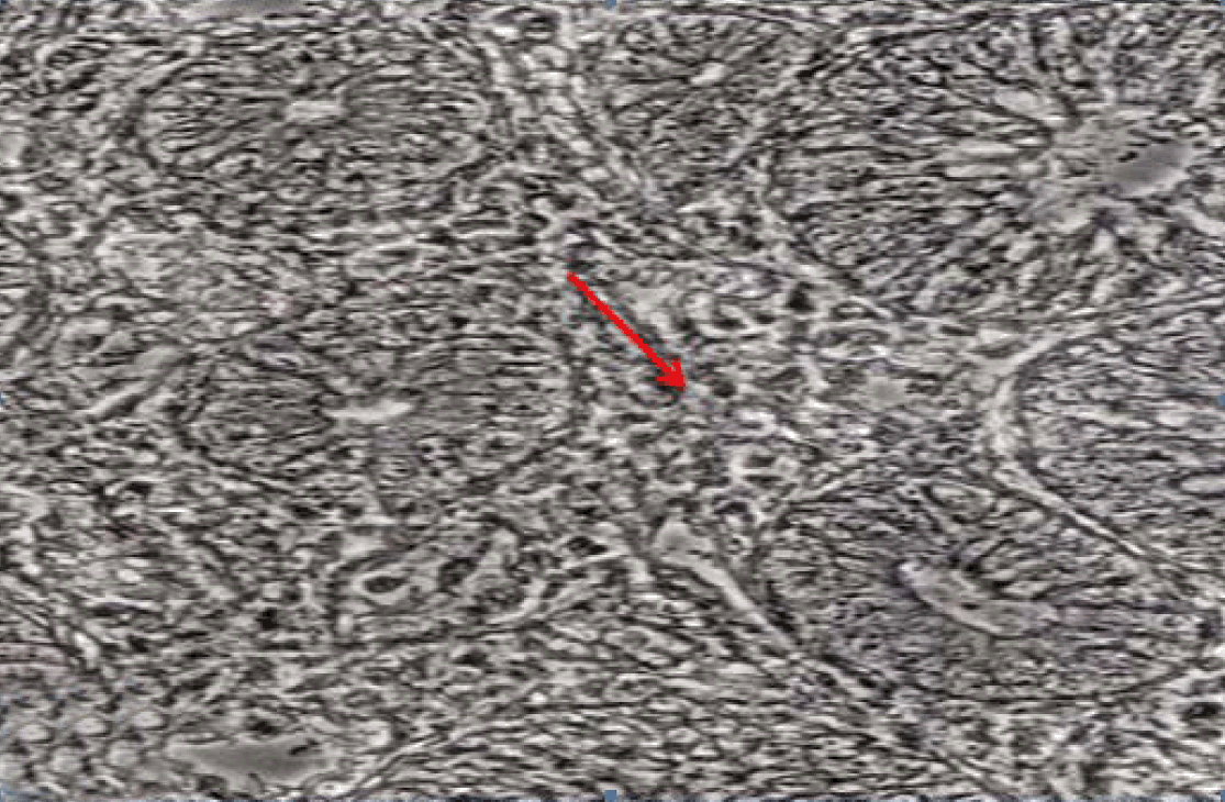
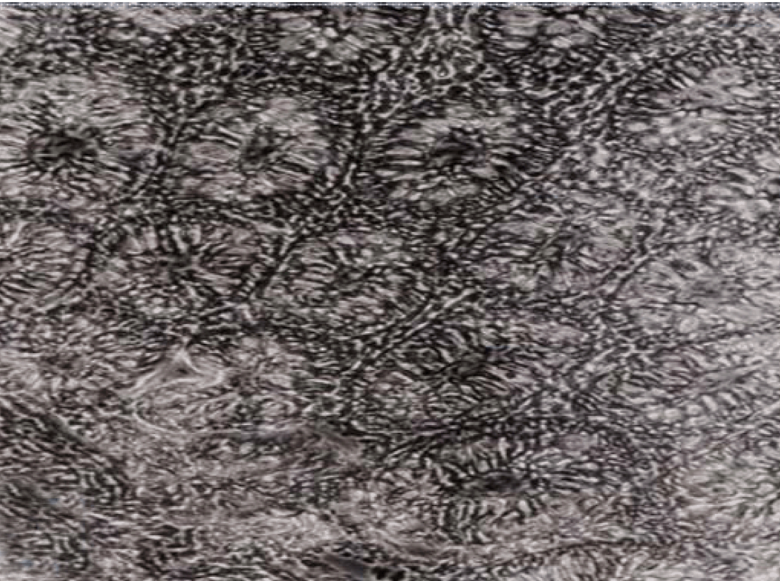
The freshly isolated jejunum cells were viable, as demonstrated by dye exclusion technique. A large number of isolated intestinal crypts and cells were seen. Cultured adult rabbit jejunum epithelial cells, two days after plating using explants technique, showed rapid epithelial growth toward the floor of culture flasks (Figure 1). The early cellular growth was characterized by being of spindle shaped cells. These cells attached closely to each other, but did not exhibit typical epithelial morphology. They proliferated most rapidly from days 2 to 3 and relatively slowly from days 4 to 5 with evidence of cellular mitosis (Figure 2). Epithelial intestinal cells harvested using collagenase technique rapidly attach to the floor of culture flasks. A large number of isolated intestinal crypts and cells were seen (Figure 3). Both collagenase and explants harvested intestinal cells rapidly converted into fibroblasts-like cells within 8-10 days in culture (Figure 4).

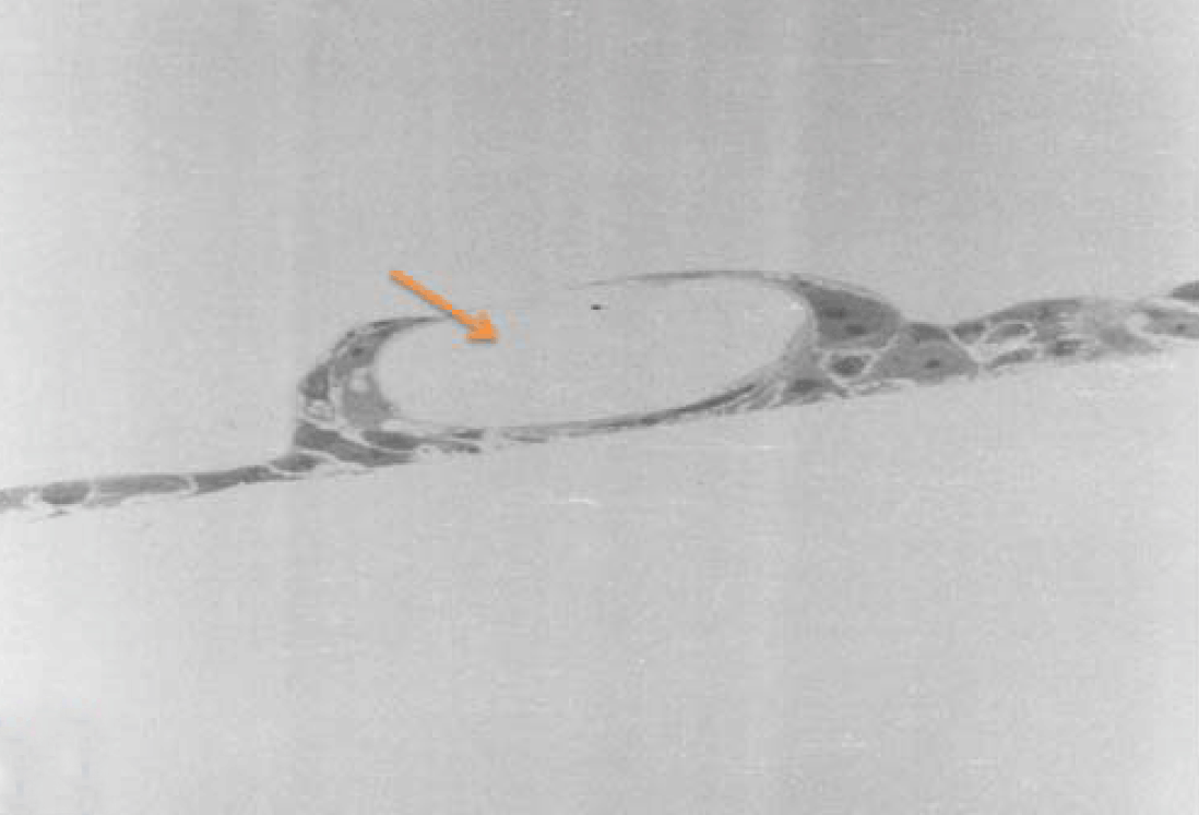
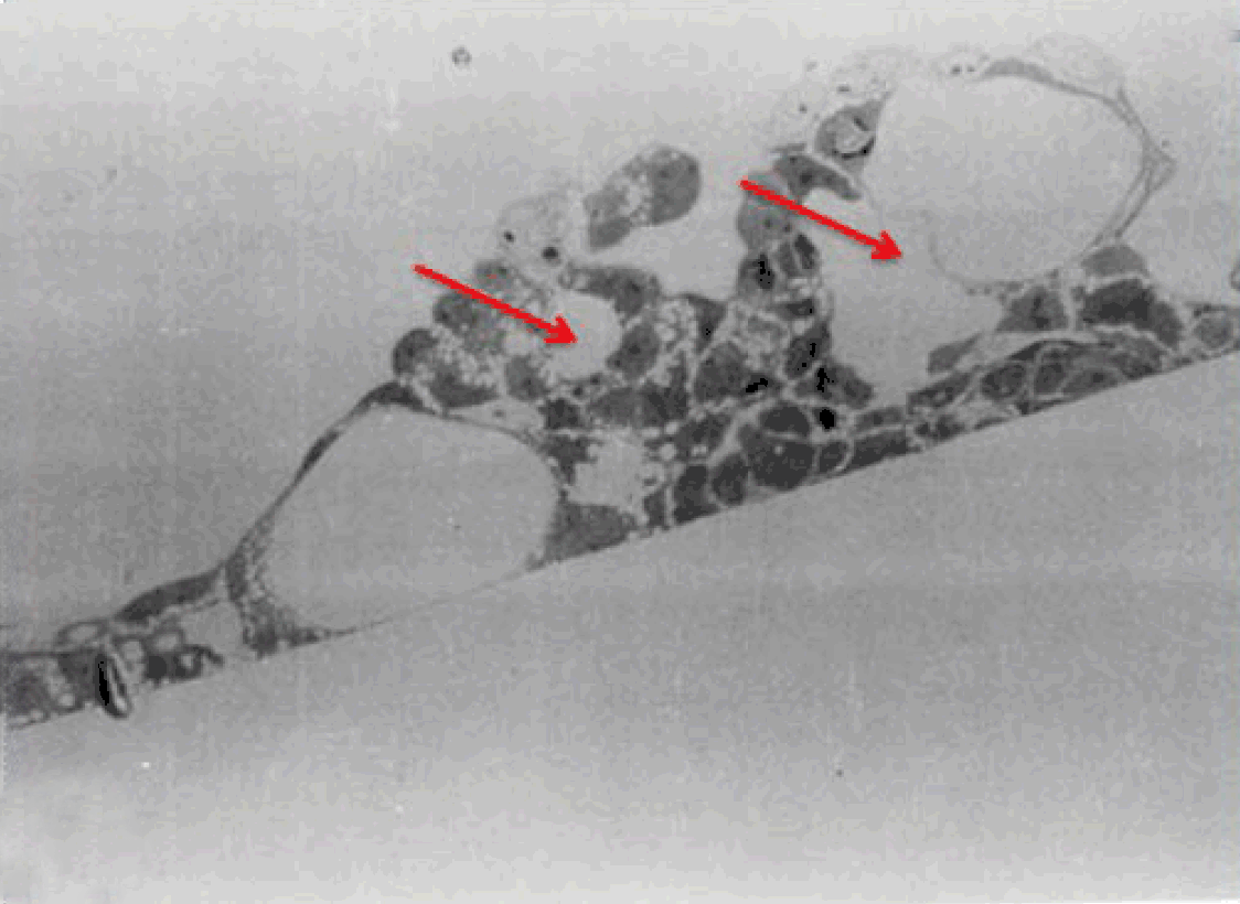
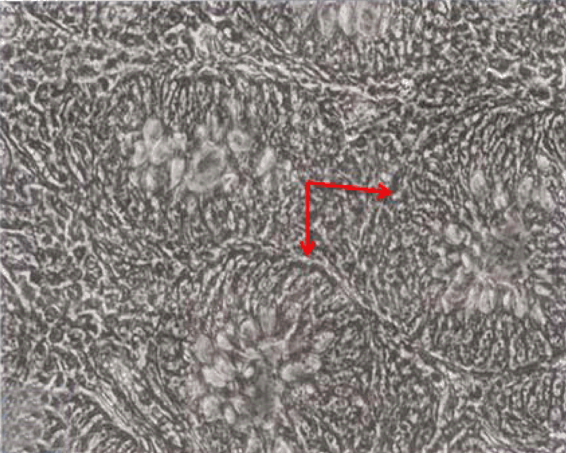
The non-polar human Caco-2 cells when grown as monolayer (Figure 5) or aggregates (Figure 6), showed no brush border. Inter-cellular cysts (ICC), acini-like structures, were occasionally observed within Caco-2 cells aggregates (Figure 6). ICCs were simple in early culture (Figure 7) but complex in later culture (18 days culture) (Figure 8). The Caco-2 cells gradually started to form increased number of rounded crypts-like structures. The early crypts were large and rounded at 21 days culture (Figure 9). Some were irregular and elongated (Figure 10) then became rounded. Few free cells remained in between such crypts (Figure 11). With the advancement of culture to nearly 4 weeks, the Caco-2 cells formed small nearly regular crypts (Figure 12). Such change is an indication for their spontaneous enterocytic differentiation, which is characterized with polarization of the cell layer with the formation of domes that increased in the density in older culture.

Transmission electron microscopy (TEM)
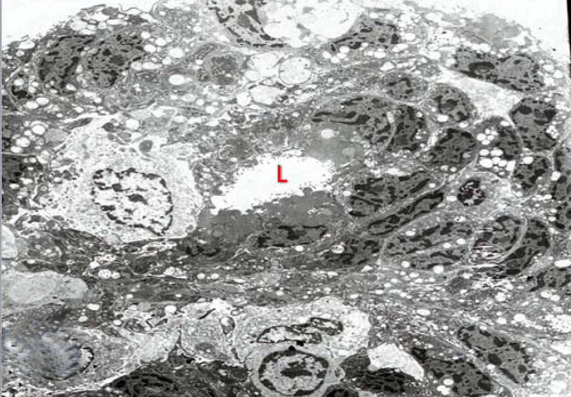
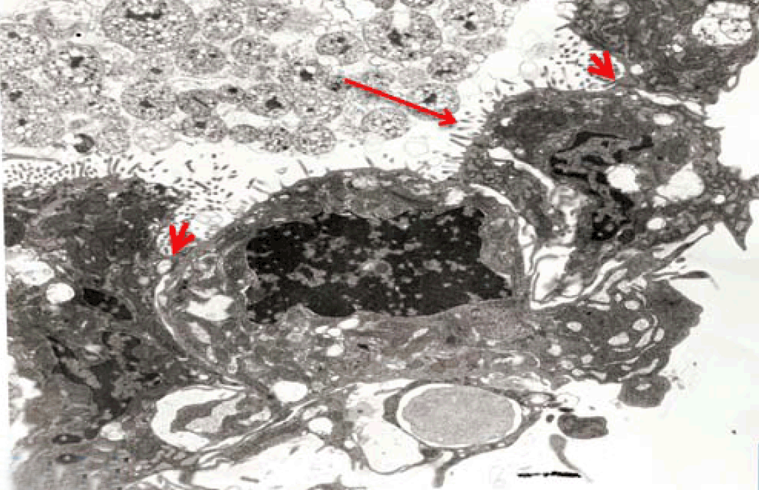
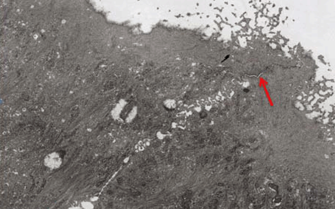
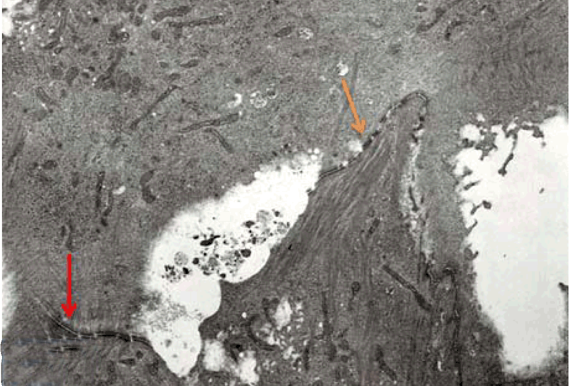
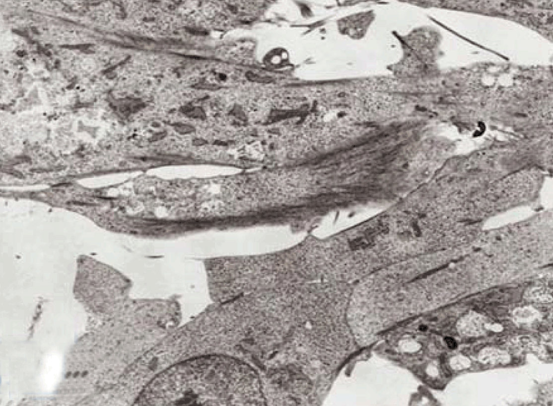
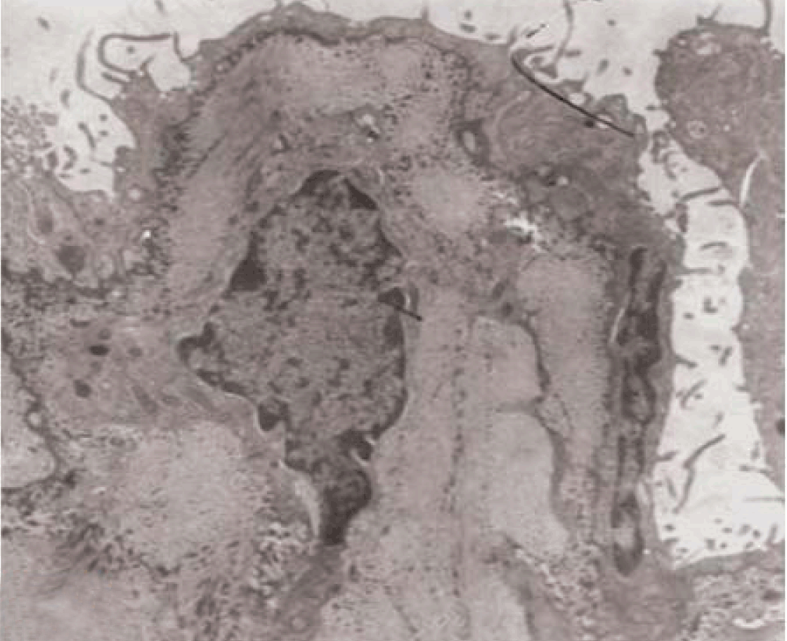
Intestinal epithelial cells on 1-2 days in culture, particularly those isolated by collagenase harvesting technique, formed simple cuboidal to columnar epithelia with basally-located nuclei. Ultrastructurally, the epithelial cells appeared closely attached to each other (Figure 13). Intestinal epithelial cells are connected by tight junctional complexes and desmosomes at the subluminal region. The intestinal cells are relatively homogeneous. Numerous long microvilli were projecting on the luminal surface, while their basal surface showed cytoplasmic projections (Figure 14). Thus, the intestinal epithelial cells in primary culture retained some characteristics of absorptive epithelial cells. Gradually the epithelial cells losses their microvilli, but retains their junctional complexes and the desmosomes (Figures 15 and 16). Deterioration of cultured epithelial cells were seen starting on day 5 (Figure 4) and the cells were converted into fibroblastic like cells on day 21 (Figure 17).

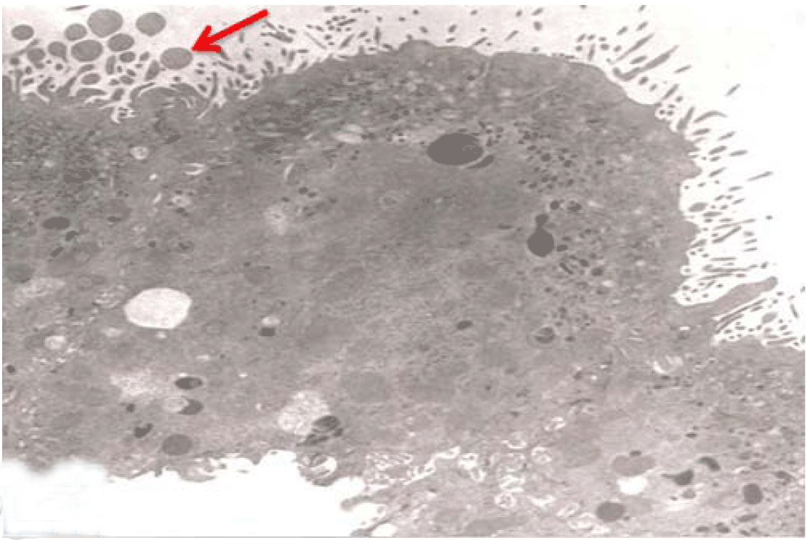
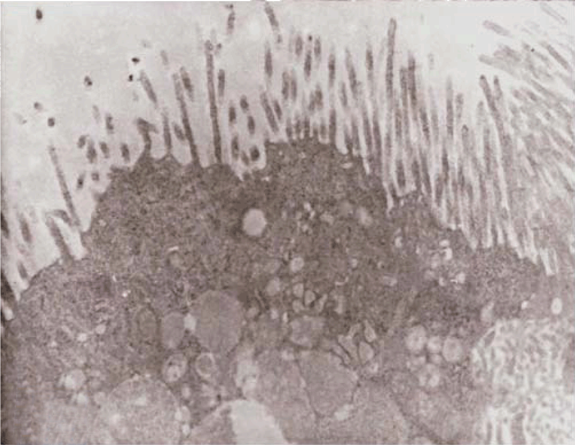
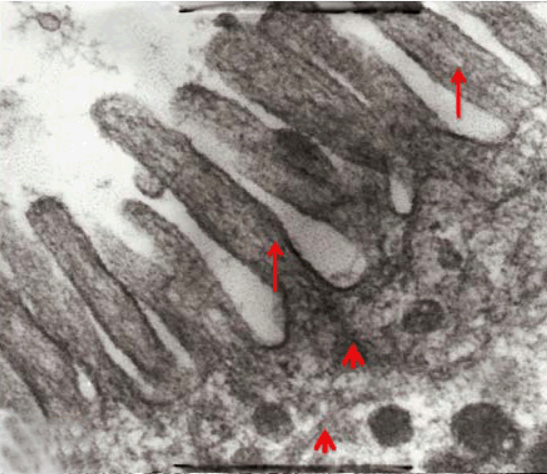
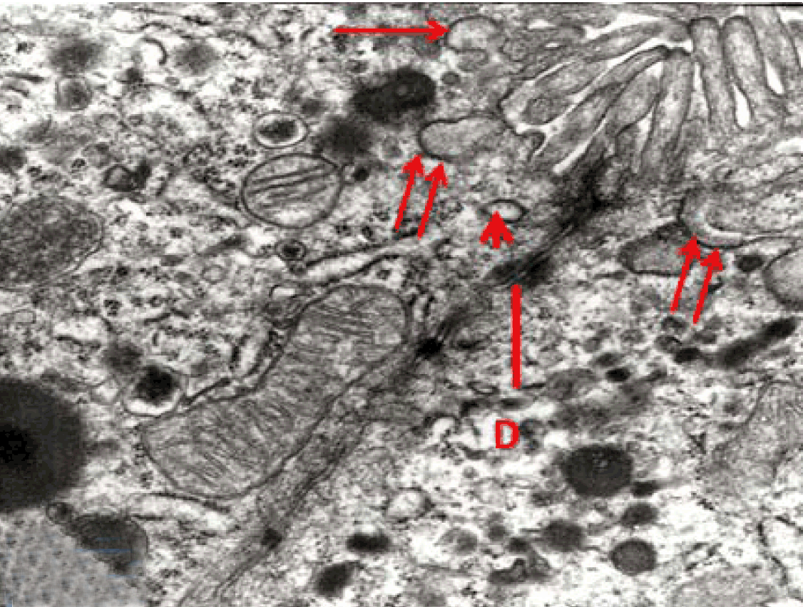
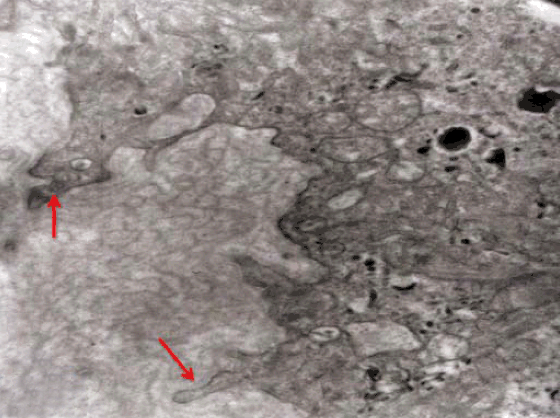
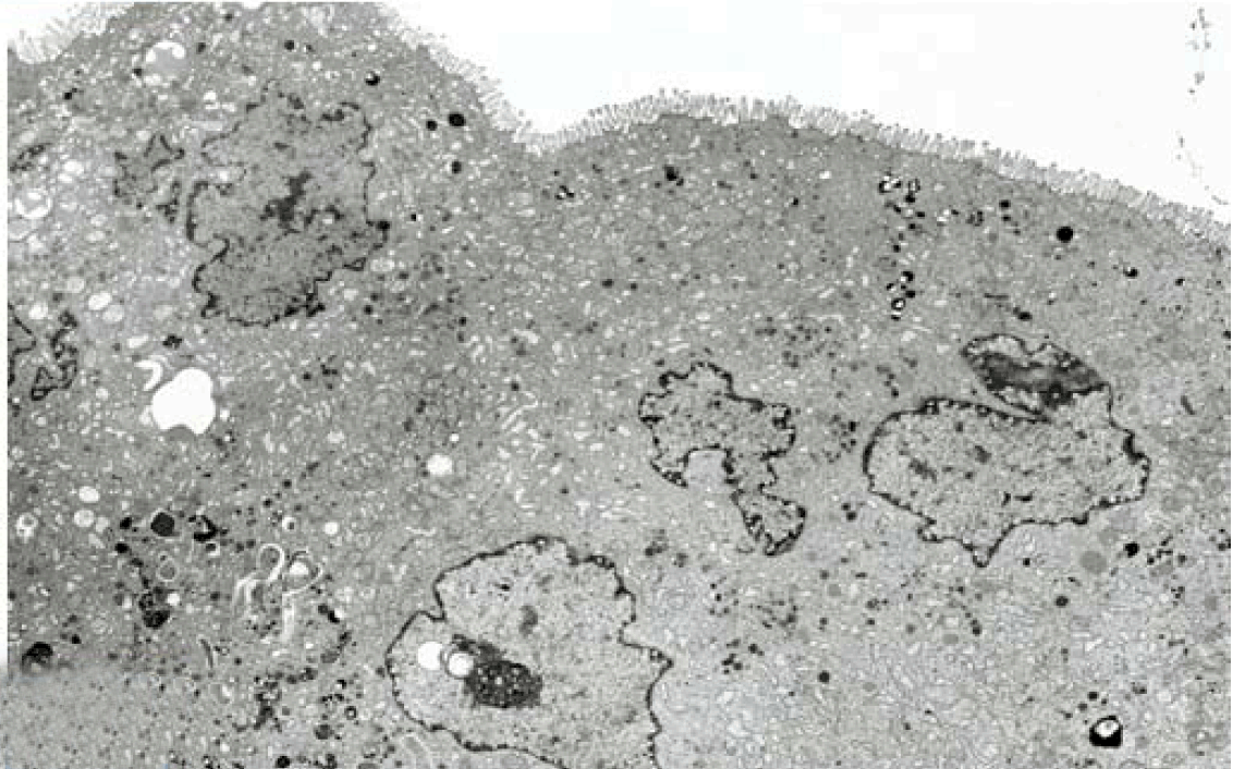
Fully polarize differentiated Caco-2 cells formed a simple cuboidal to columnar epithelium with basally-located nuclei (Figure 18). Concurrent with monolayer formation an increase in cell height, sparse microvilli with a few actin core filaments was seen. Early surfaced microvilli were short, irregular with blebs-like projections (Figure 19) then the villi became gradually lengthened and increased in density (Figure 20). Gradually the actin core bundles extended into the subjacent cytoplasm, forming the characteristic terminal web in fully differentiated Caco-2 cells (Figure 21). The cell height and microvilli- density continued to increase throughout the culture period. Coated pits and vesicles, apical tubo-vesicular system was also seen (Figures 21 and 22). Fully differentiated Caco-2 cells maintained their junctional complexes and the desmosomes. Moreover few basal projections were commonly seen (Figure 23). Frequent syncytial like growth pattern formation of fully polarize differentiated Caco-2 cells was seen with bizarre nuclear shape and numerous regular dense surface microvilli at 25 days culture (Figure 24).

Scanning electron microscopy (SEM)
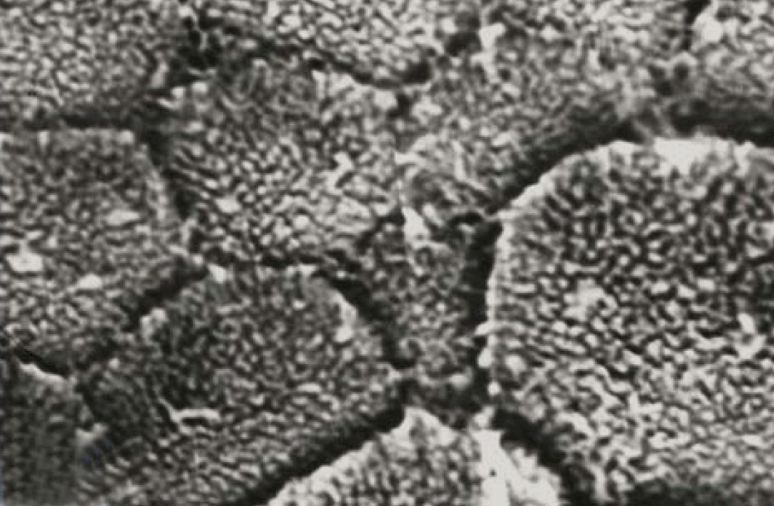
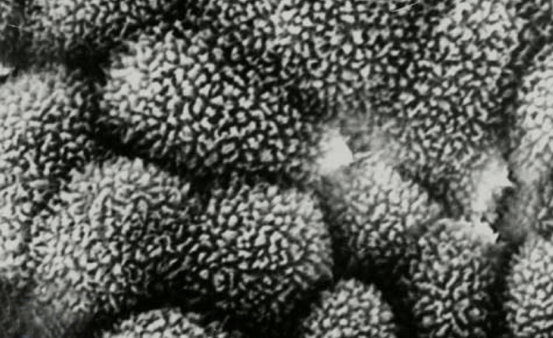
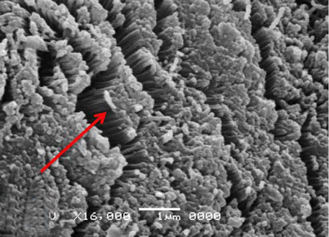
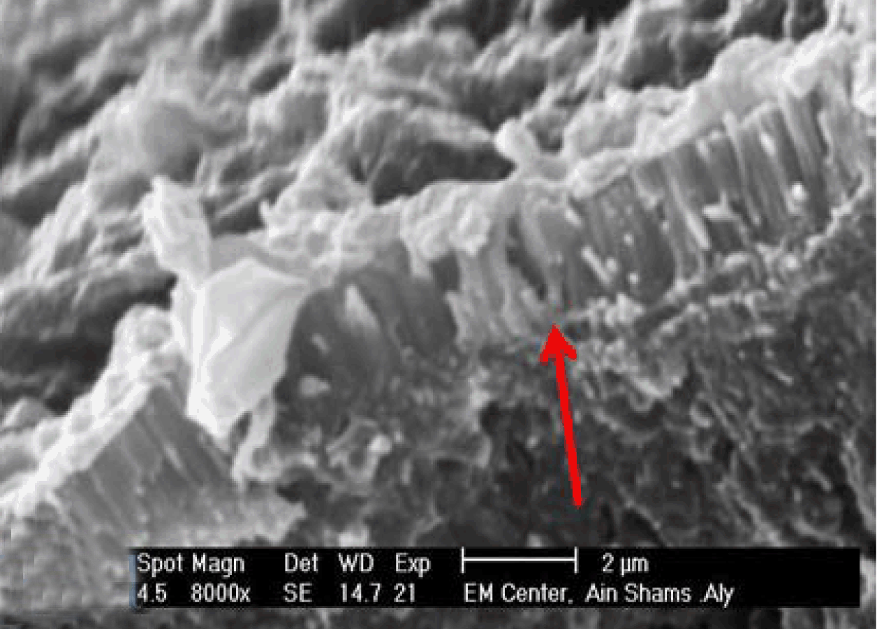
Intermediately differentiated Caco-2 cells are nearly polygonal shaped cells and the surfaces are flat and bearing short microvilli (Figure 25). While fully differentiated Caco-2 cells are polygonal, with dome-shaped surfaces bearing high density surface microvilli (Figure 26). This is quite similar to the normal phenotype surface microvilli which appeared in the brush border of the jejunal epithelial cells of the normal rabbit (Figure 27). The jejunal epithelium showed also well-established epithelial junctional complexes including surface zonula occludes (tight junction), around the apical membrane (Figure 28). Fully differentiated Caco-2 cells showed surface microvilli (brush border) and well established apical epithelial junctional complexes (Figure 29).

Immunofluorescence
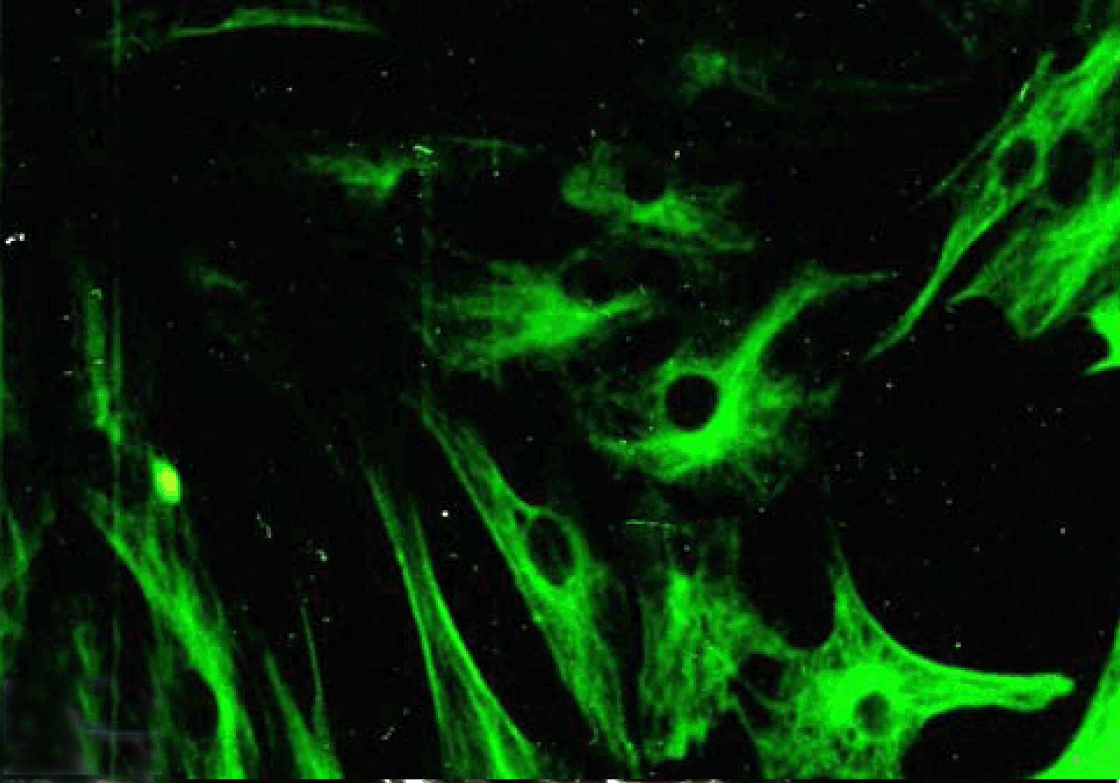
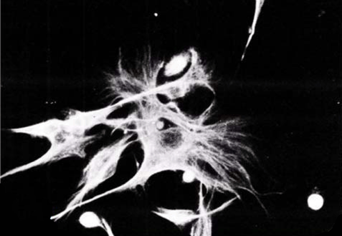
The cultured adult rabbit jejunum epithelial cells expressed cytokeratin on days 1 to 5 in culture, suggesting that most cells were epithelial in nature (Figure 30). Both collagenase and explants harvested intestinal cells rapidly converted into fibroblast like cells within 8-10 days in culture and failed to express the characteristic cytokeratin. Cultured Caco-2 cells expressed the characteristic cytokeratin labeling confirming their epithelial origin (Figure 31). Pancytokeratin was expressed in the perinuclear vicinity as filaments that are radiating towards the cell periphery.

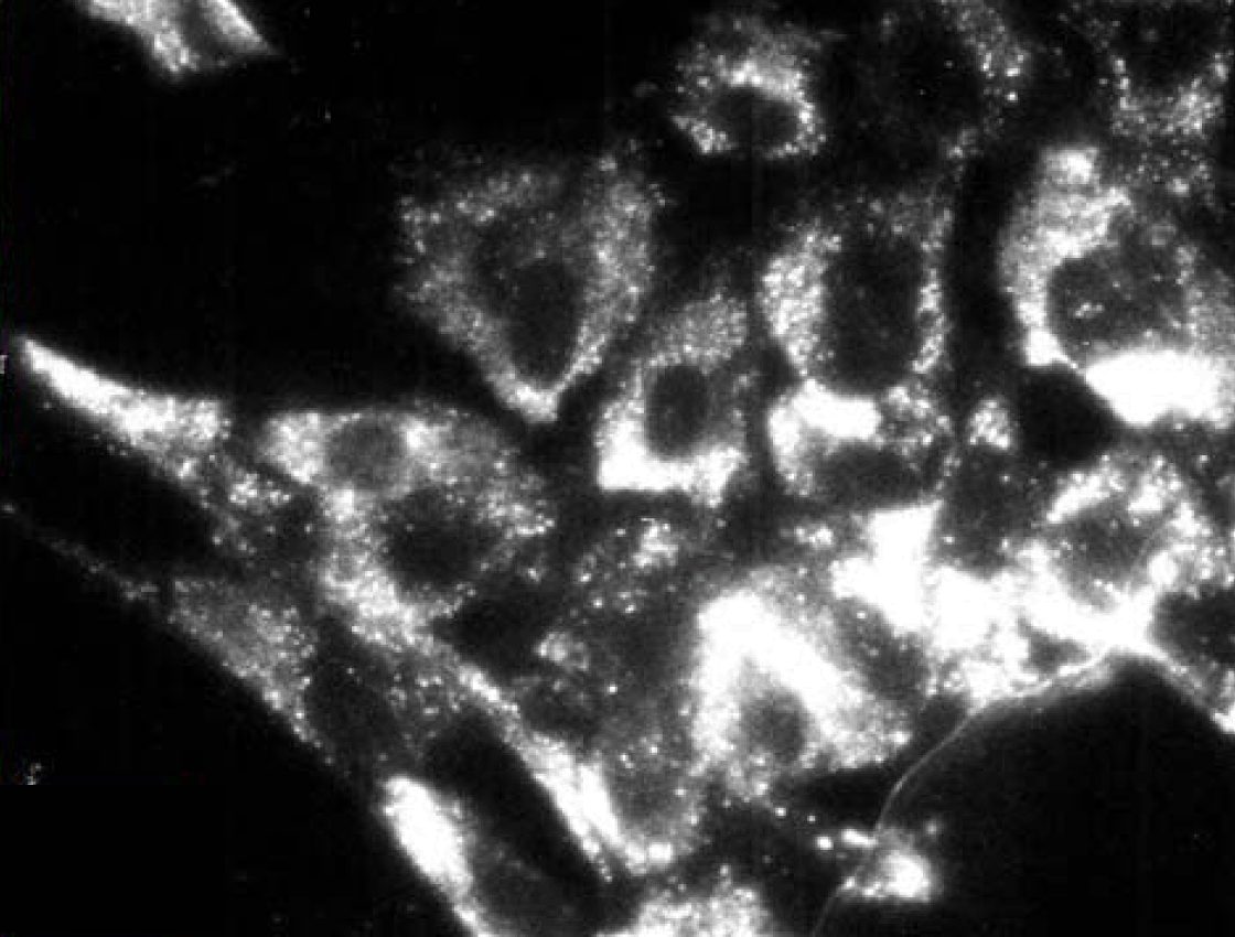
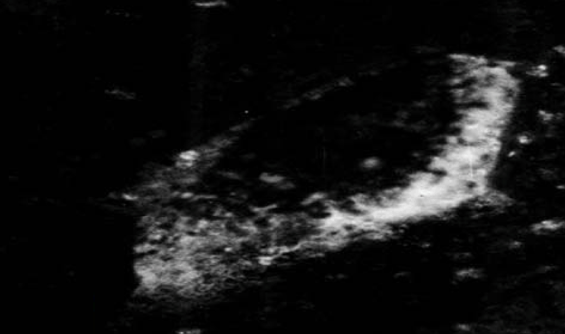
Polarized Caco-2 cell monolayer in culture stained positive with monoclonal antibody HBB specific for sucrase-isomaltase as isolated cells (Figure 32) or as aggregated acini (Figure 33).

Immunoelectron microscopy
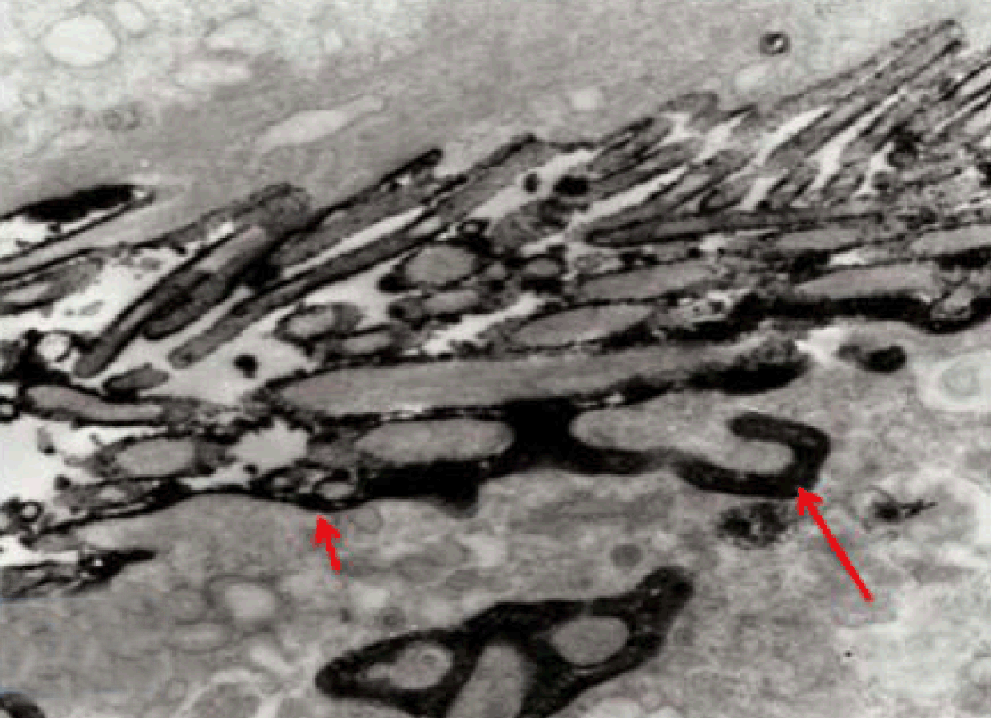
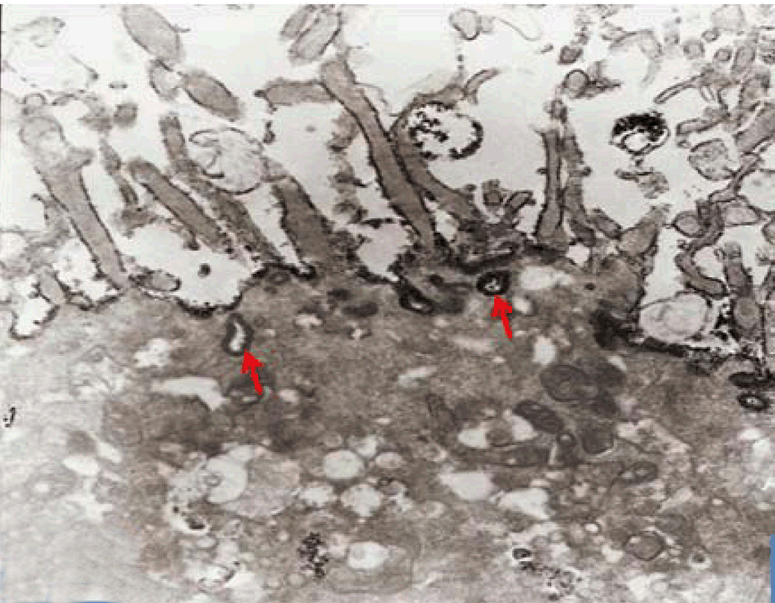
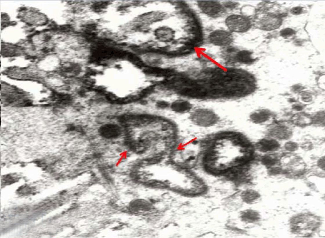
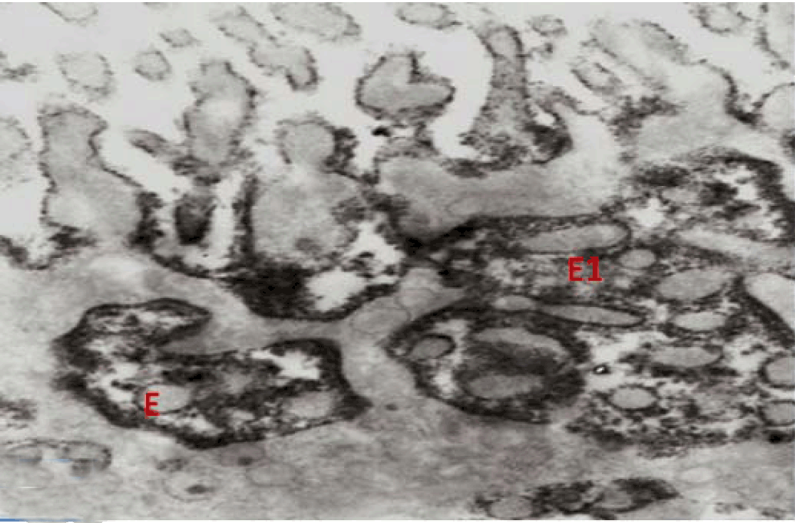
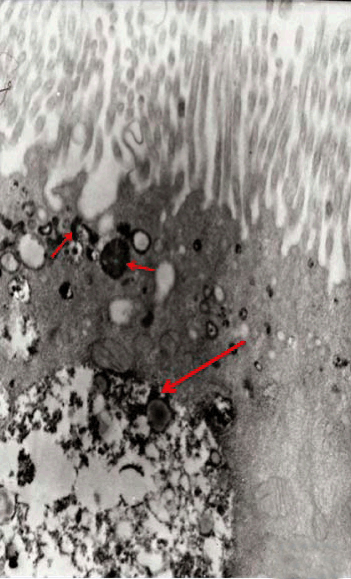
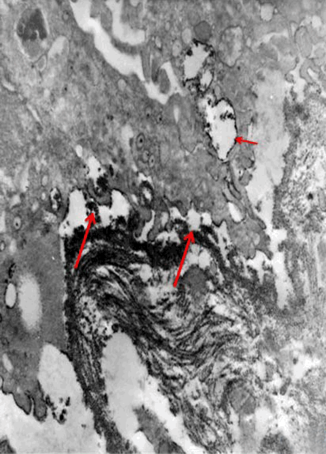
Apical endocytic pathways in polarized Caco-2 cells were investigated by following the uptake and transcytosis of Goat anti-rabbit IgG that was conjugated to horseradish peroxidase (IgG-HRP). The uptake was seen at the surface of microvilli and at the apical cell invagination or pit (caveolae) (Figure 34). Coated pits are frequently observed on the apical membrane and at the base of the microvilli in polarized Caco-2 cells monolayer. Some small apical endosomes were labeled (Figure 35). Also tubo-vesicular systems were labeled (Figure 36). After 15 minutes, larger endosomes became labeled; some were seen connected to the surface membrane (Figure 37). By 30 minutes, vacuolar endosomes including multivesicular bodies were presented deeper in the cell and labeled elements were widespread in the apical cytoplasm (Figure 38). No labeled elements occurred in the basal regions, below the nucleus. Also no labeling was seen in the surface of microvilli and in the apical coated caveolae. With longer IgG-conjugated-HRP incubations, 60 minutes, the penetration of label elements became very extensive reaching the basal region of the Caco-2 cells (Figure 39). Finally IgG-conjugated-HRP was seen deep to the cell base (Figure 39). Such labeling is an evidence for a receptor-mediated transcytotic pathway from the apical surface to the basolateral border of Caco-2 cells.


Cursor on image to zoom/Click text to open image
Figure 1: Phase contrast micrograph of normal adult rabbit jejunum (explants isolation) showing epithelial cell growth toward the floor of cultured flasks. The cellular growth was characterized by spindle shaped cells. (E) explants (2 days culture), x200.


Cursor on image to zoom/Click text to open image
Figure 2: Phase contrast micrograph of normal adult rabbit jejunum (explants isolation) showing epithelial cells attached closely to each other in monolayer culture but not exhibiting typical epithelial morphology (cobblestone appearance). Note evident of mitosis (arrow). (E) explants (3 days culture), x200.


Cursor on image to zoom/Click text to open image
Figure 3: Phase contrast micrograph of normal adult rabbit jejunum (collagenase isolation) showing epithelial cells attached to the floor of the culture flask. Note the large number of isolated intestinal crypts and cells in-between, x600.


Cursor on image to zoom/Click text to open image
Figure 4: Phase contrast micrograph of normal adult rabbit jejunum (collagenase isolation) showing that intestinal cells rapidly converted into fibroblasts-like cells within 8–10 days in culture, x200.


Cursor on image to zoom/Click text to open image
Figure 5: Phase contrast micrograph of Caco-2 cells in early culture showing that Caco-2 cells are grown as monolayer (12 days culture), x400.


Cursor on image to zoom/Click text to open image
Figure 6: Phase contrast micrograph of Caco-2 cells in early culture showing that Caco-2 cells are grown in aggregates, with no brush border (14 days culture), x400.


Cursor on image to zoom/Click text to open image
Figure 7: Micrograph of semi thin section of Caco-2 cells in early culture, showing that Caco-2 cells are forming simple inter-cellular cysts (acini-like structures) (arrow) within the Caco-2 aggregates (14 days culture), x200.


Cursor on image to zoom/Click text to open image
Figure 8: Micrograph of semi thin section of Caco-2 cells in early culture showing, the complex type of Inter-cellular cysts (acini-like structures) (arrow) within the Caco-2 aggregates (18 days culture), x200.


Cursor on image to zoom/Click text to open image
Figure 9: Phase contrast micrograph of Caco-2 cells showing that the early crypts were large and rounded (arrow) (21 days culture), x600.


Cursor on image to zoom/Click text to open image
Figure 10: Phase contrast micrograph of Caco-2 cells showing that some of the crypts are irregularly elongated (arrow) (21 days culture), x600.


Cursor on image to zoom/Click text to open image
Figure 11: Phase contrast micrograph of Caco-2 cells showing free cells in between the crypts (arrow) (22 days culture), x600.


Cursor on image to zoom/Click text to open image
Figure 12: Phase contrast micrograph of Caco-2 cells showing Caco-2 cells forming small nearly regular and rounded crypts. Note that the free cells in-between the crypts decreased in density and the crypts became smaller and numerous (23 days culture), x600.


Cursor on image to zoom/Click text to open image
Figure 13: An electron micrograph of normal adult rabbit jejunal epithelial cells (collagenase harvested) in early culture (3 days), showing an intestinal crypt. (L) Lumen, x10,000.


Cursor on image to zoom/Click text to open image
Figure 14: An electron micrograph of normal adult rabbit jejunal epithelial cells (collagenase harvested), showing that Intestinal epithelial cells are connected by tight junctional complexes at the subluminal region (arrow head) with numerous microvilli projecting on the luminal surface (long arrow), while their basal surface showed cytoplasmic projections. Note those intestinal epithelial cells are formed of simple cuboidal to columnar epithelia with basally-located nuclei (5 days culture), x12,000.


Cursor on image to zoom/Click text to open image
Figure 15: An electron micrograph of normal adult rabbit jejuna epithelial cells (collagenase harvested), showing that the cells lost their microvilli and the apical granules within 8–10 days in culture, yet retained their junctional complexes (arrow). x17,000.


Cursor on image to zoom/Click text to open image
Figure 16: An electron micrograph of normal adult rabbit jejunum epithelial cells (collagenase harvested) showing that the cells are retaining their junctional complexes and desmosomes (arrow) at 12 days in culture, x17,000.


Cursor on image to zoom/Click text to open image
Figure 17: An electron micrograph of normal adult rabbit jejuna epithelial cells (collagenase harvested) in older culture (21 day culture) showing that the cells were converted into fibroblastic like cells, x17,000.


Cursor on image to zoom/Click text to open image
Figure 18: An electron micrograph of Caco-2 cells showing simple cuboidal to columnar epithelial cells with basally-located nuclei (18 days culture), x17,000.


Cursor on image to zoom/Click text to open image
Figure 19: An electron micrograph of Caco-2 cells in early culture showing that early surfaced microvilli are short, irregular, note the blebs-like structures (arrow) (21 days culture), x17,000.


Cursor on image to zoom/Click text to open image
Figure 20: An electron micrograph of Caco-2 cells showing that the surface microvilli increased in length and in density at 23 days culture, x17,000.


Cursor on image to zoom/Click text to open image
Figure 21: An electron micrograph of fully differentiated polarized Caco-2 cells showing that the actin core bundles have extended into the subjacent cytoplasm (arrows), forming the characteristic terminal web (arrow heads), x40,000.


Cursor on image to zoom/Click text to open image
Figure 22: An electron micrograph of fully differentiated polarized Caco-2 cells showing apical coated pits (arrow), vesicles (arrow head) and apical tubo-vesicular system (double arrow). Note that Caco-2 cells maintained their junctional complexes and the desmosomes (D), x30,000.


Cursor on image to zoom/Click text to open image
Figure 23: An electron micrograph of fully differentiated polarized Caco-2 cells showing a few basal projections (arrow). x25,000.


Cursor on image to zoom/Click text to open image
Figure 24: An electron micrograph of some fully differentiated polarized Caco-2 cells exhibiting syncytium like structural pattern with bizarre nuclear shape and numerous regular dense surface microvilli (25 days culture), x25,000.


Cursor on image to zoom/Click text to open image
Figure 25: A scanning electron micrograph of fully differentiated Caco-2 cells showing that the cells are nearly polygonal and the surfaces are flat and bearing short microvilli (21 days culture), x4,000.


Cursor on image to zoom/Click text to open image
Figure 26: A scanning electron micrograph of fully differentiated Caco-2 cells. The cells have polygonal dome-shaped surfaces bearing high density surface microvilli (23 days culture), x4,000.


Cursor on image to zoom/Click text to open image
Figure 27: A scanning electron micrograph of normal adult rabbit jejunal mucosal cells showing normal phenotype surface microvilli. The brush border is formed of tightly packed long microvilli (arrow) arranged in a highly even pattern of closely and densely packed rows with their axes oriented perpendicular to the long axis of their cells( 4 days culture), x16,000.


Cursor on image to zoom/Click text to open image
Figure 28: A scanning electron micrograph of normal adult rabbit jejunal mucosal cells in cross section view showing normal phenotype surface microvilli (brush border) and well established epithelial junctional complexes including surface zonula occludes (tight junction) (long arrow) around the apical membrane (4 days culture), x8,000.


Cursor on image to zoom/Click text to open image
Figure 29: A scanning electron micrograph of fully differentiated Caco-2 cells showing surface microvilli (brush border) and well established apical epithelial junctional complexes (long arrow) (23 days culture), x8,000.


Cursor on image to zoom/Click text to open image
Figure 30: Immunofluorescence micrograph of early cultured adult rabbit jejunal mucosal cells showing the characteristic cytokeratin distribution. Pancytokeratin is seen in the perinuclear region as filaments that are radiating toward the cell periphery, x400.


Cursor on image to zoom/Click text to open image
Figure 31: Immunofluorescence micrograph of Caco-2 cells stained for cytokeratin. Pancytokeratin is expressed in the perinuclear and paranuclear vicinity as the intermediate filaments are seen radiating toward the cell periphery. Note the variability in cells size, x400.


Cursor on image to zoom/Click text to open image
Figure 32: Immunofluorescence micrograph of differentiated polarized Caco-2 cells in early culture showing the characteristic immune-localization of sucrase-isomaltase Note the labeling pattern (no counter stain was used), x400.


Cursor on image to zoom/Click text to open image
Figure 33: Immunofluorescence micrograph of polarized Caco-2 cells in late culture showing the characteristic immune localization of sucrase-isomaltase within well differentiated Caco-2 intestinal acini. Note the labeling pattern (no counter stain was used) (23 days culture), x1,000.


Cursor on image to zoom/Click text to open image
Figure 34: Immunoelectron micrograph of unstained polarized Caco-2 cells monolayer in culture showing the apical uptake of Goat anti-rabbit IgG conjugated to horseradish peroxidase (IgG-HRP). Note the extensive labeling at the surface of microvilli and at the apical cell invagination or pit caveolae (arrow) at the base of the microvilli. Note the apical tubular system (long arrow) (23 days culture), x20,000.


Cursor on image to zoom/Click text to open image
Figure 35: Immunoelectron micrograph of unstained polarized Caco-2 cells monolayer in culture showing the apical uptake of Goat anti-rabbit IgG conjugated to horseradish peroxidase (IgG-HRP). Note the labeling at small apical endosomes at the base of the microvilli (arrow) (23 days culture), x16,000.


Cursor on image to zoom/Click text to open image
Figure 36: Immunoelectron micrograph of unstained polarized Caco-2 cells monolayer in culture showing the apical uptake of Goat anti-rabbit IgG conjugated to horseradish peroxidase (IgG-HRP). Note the labeling at the tubo-vesicular system (short arrow) as well as coated caveolae (long arrow) (23 days culture), x30,000.


Cursor on image to zoom/Click text to open image
Figure 37: Immunoelectron micrograph of unstained polarized Caco-2 cells monolayer in culture showing the apical uptake of Goat anti-rabbit IgG conjugated to horseradish peroxidase (IgG-HRP). Note the labeling of larger endosomes (E); some were seen connected to the surface membrane at the base of microvilli (E1) (23 days culture), x30,000.


Cursor on image to zoom/Click text to open image
Figure 38: Immunoelectron micrograph of unstained polarized Caco-2 cells monolayer in culture showing the apical uptake of Goat anti-rabbit IgG conjugated to horseradish peroxidase (IgG-HRP). Note the labeling of large vacuolar endosomes that were seen deeper in the cell (Long arrow). Note also that the labeled elements were widespread in the apical cytoplasm (Short arrow) (23 days culture), x30,000.


Cursor on image to zoom/Click text to open image
Figure 39: Immunoelectron micrograph of unstained polarized Caco-2 cells monolayer in culture showing the apical uptake of Goat anti-rabbit IgG conjugated to horseradish peroxidase (IgG-HRP) (60 minutes) showing the penetration of label elements at the basal region of the Caco-2 cells (short arrow) as well as beneath the basal region (long arrow) (23 days culture). x40,000.


DISCUSSION

In the present study, an attempt was made to establish long term culture model of adult rabbit normal isolated intestinal epithelial cells. However, it was found that the isolated intestinal cells rapidly lost their characteristic phenotype and converted into fibroblast-like cells within one week in culture. Similar results were previously reported [2] and denote that contamination with fibroblasts led to failure of such culture model. Moreover, the mouse jejunum, when incubated for four hours, exhibited morphological deterioration of the villi with denudation of the epithelia while it retained cAMP-induced potential difference (PD) [17]. Therefore, we described an alternating in vitro cell culture system consisting of a monolayer of viable, polarized and spontaneously fully differentiated Caco-2 cells that were similar to that found in the small intestine in the absence of inducers of differentiation.

In the present work, under the standard culture conditions, Caco-2 cells underwent spontaneously enterocytic differentiation both ultrastructurally and immunocytochemically. The cells developed junctional complexes, with desmosomes, very similar to adult differentiated intestinal epithelial cells. The above mentioned observations are in accordance with Hilgers et al. [11] who stated that Caco-2 cells spontaneously differentiate in culture to form confluent monolayers which both structurally and functionally resemble the small intestinal epithelium. Caco-2 cells have been used by other authors in regard to a broad spectrum of intestinal and epithelial parameters [18].

The brush border on the apical surface of enterocytes is a highly specialized structure, well-adapted for efficient digestion and nutrient transport, whilst at the same time providing a protective barrier for the intestinal mucosa [19]. The brush border is continuous with a densely ordered array of microvilli, protrusions of the plasma membrane, which are supported by actin-based microfilaments and interacting proteins and anchored in an apical network of actomyosin and intermediate filaments, the so-called terminal web [19]. In the present study Caco-2 cells in late passage of 30–40 expressed sucrase-isomaltase (SI) characteristic of epithelial enterocytes. This is supported by a similar observation [14] that the major site of expression of the enzyme sucrase-isomaltase (SI) is the brush border of the small intestine. Moreover, Caco-2, have been used extensively for analyzing the expression and biosynthesis of the enzyme sucrase-isomaltase (SI) and other brush border hydrolases [15],[16],[20].

The fully differentiated Caco-2 cells were polygonal with domed shaped surfaces bearing high density surface microvilli and well established epithelial junctional complexes including surface zonula occludes (tight junction) around the apical membrane. Gradually the actin core bundles spreads into the apical villi extending into the subjacent cytoplasm, forming the characteristic terminal web in fully differentiated Caco-2. The cell height and microvilli density continued to increase throughout the culture period. Coated pits and vesicles, apical tubo-vesicular systems were also seen.

Wang et al (2017)[21] stated that dysfunction of the epithelial barrier is a hallmark of inflammatory intestinal diseases. The intestinal epithelial barrier is maintained by expression of tight junctions that connect adjacent epithelial cells and seal the para-cellular space. IL-22 is critical for the maintenance of intestinal barrier function through promoting anti-pathogen responses and regeneration of epithelial tissues in the gut utilizing polarized Caco-2 cell. In the present work, Caco-2 cells showed spontaneous differentiation in three weeks resulting in a monolayer of cells that have morphological and functional characteristics of mature enterocytes. Samak et al (2015) [10]concluded that the disruption of tight junctions and barrier dysfunction may lead to mucosal translocation of luminal toxins and pathogens to induce inflammatory process and colitis. Epithelial cells and their intercellular junctions play a primary role in the intestinal mucosal diffusion barrier to the invasion of noxious agents. Lee et al (2017) [22] indicated that Astragaloside II (AS II) can contribute to epithelial barrier repair following intestinal injury, and may offer a therapeutic avenue in treating irritable bowel disease in Caco-2 cells culture model.

In the present study the uptake IgG-HRP was seen at the apical cell invagination caveolae that were frequently observed on the apical membrane at the base of the microvilli in polarized monolayer of Caco-2 cells. After 15 minutes, larger endosomes became labeled and by 30 minutes vacuolar endosomes including multivesicular bodies present deeper in the cell. After 40 min, labeled elements were widespread in the apical cytoplasm. No labeled elements occurred in the basal regions, below the nucleus. After 60 minutes, the penetration of labeled elements became very extensive reaching the basal region of the Caco-2 cells. Finally IgG-HRP was seen deep in the cell base. Such labeling is an evidence for a receptor-mediated transcytotic pathway from the apical surface to the basolateral border of Caco-2 cells. Receptor-mediated endocytosis is a specific process for cells to take up small and large molecular legends, including hormones, growth factors, enzymes, and plasma proteins [23]. Thus Receptor-mediated endocytosis, which was demonstrated in Caco-2 culture model, could be utilized for efficient drug delivery to the target cells with high expression of the receptors. For examples, transferrin receptor (TfR) and insulin receptor (IR) mediated endocytosis systems have been used for small molecules and therapeutic protein delivery [24]. Besides transport of macromolecules, endocytosis is also involved in antigen presentation, maintenance of cell polarity and regulation of cell-surface receptor expression [8].

Receptor-mediated endocytosis (RME) is driven by clathrin-coated vesicle (CCV) cycle [25]. This cycle begins with the assembly of the coat proteins; clathrin, into coated pits of apical cell membrane (ACM). Clathrin assembles into a lattice-like structure and induces inward curvature of the pit as it grows with the successive addition of coat proteins. The coated pit invaginates and eventually detaches from the ACM, forming a coated vesicle [26]. Clathrin-mediated endocytosis is the best characterized endocytic mechanism and is the predominant pathway for macromolecule uptake along epithelia [6]. Broecket al (2007) [27] suggested the involvement of the clathrin-mediated endocytic internalization route in the uptake of cholera, utilizing human intestinal Caco-2 cells. Receptor-mediated endocytosis depends on the integrity of the actin cytoskeleton and the microtubules [6].

In addition to clathrin- and caveolae-mediated endocytosis, clathrin and caveolae-independent endocytosis exists. One example of the independent endocytosis is the internalization of human IgG in Caco-2 cells. In order to understand the mechanism of the absorption of therapeutic monoclonal antibodies by the human epithelial cells, the endocytosis and internalization of a human IgG into Caco-2 was examined in the present work. Xiao and Gan (2013) [23] found that endocytosis of the human IgG into Caco-2 cells was pH, temperature, and ATP-dependent. In addition, caveolae-dependent endocytosis inhibitors, Nystatin and Indomethacin, had no significant effects on the cell association and binding of human IgG to the Caco-2 cells, indicating that the internalization is a clathrin- and caveolae-independent endocytosis [23]. Yuan et al (2017) [28] studied transport of isorhapontigenin (ISO) across Caco-2 cells and found that ISO has anti-inflammatory effect, anti-oxidation effect and anti-cancer effect. The results suggested that transport mode of ISO was mainly passive diffusion in Caco-2 cell models, and P-gp and MRP may be involved in the transport of ISO.


CONCLUSION

Long term culture model of adult rabbit normal isolated intestinal epithelial cells (explants or collagenase harvested) was unstable model for intestinal barrier experiments. Caco-2 cells model could be a significant model for research on drug delivery across the intestinal epithelium. It can also be used to evaluate strategies for investigating and/or enhancing absorption, macromolecular transport by different ways across the intestinal epithelium; such as passive transcellular, independent endocytosis and carrier receptor-mediated endocytosis mechanisms. Future research is required to further characterize different active cellular efflux pumps and transporters across the enterocytes and Caco-2 cells model.


REFERENCES
  1. Capdeville Y, Frézal J, Jos J, Rey J, Lamy M. Culture of calf intestinal tissue: Study of cell differentiation and disaccharase activities. [Article in French]. C R Acad Sci Hebd Seances Acad Sci D 1967 Jan 16;264(3):519–21.   [PubMed]    Back to citation no. 1
  2. Kedinger M, Haffen K, Simon-Assmann P. Intestinal tissue and cell cultures. Differentiation 1987;36(1):71–85.   [CrossRef]   [PubMed]    Back to citation no. 2
  3. Mostov KE, Simister NE. Transcytosis. Cell 1985 Dec;43(2 Pt 1):389–90.   [PubMed]    Back to citation no. 3
  4. Mostov K, Apodaca G, Aroeti B, Okamoto C. Plasma membrane protein sorting in polarized epithelial cells. J Cell Biol 1992 Feb;116(3):577–83.   [CrossRef]   [PubMed]    Back to citation no. 4
  5. Zacchi P, Stenmark H, Parton RG, et al. Rab17 regulates membrane trafficking through apical recycling endosomes in polarized epithelial cells. J Cell Biol 1998 Mar 9;140(5):1039–53.   [CrossRef]   [PubMed]    Back to citation no. 5
  6. Gekle M, Freudinger R, Mildenberger S. Inhibition of Na+-H+ exchanger-3 interferes with apical receptor-mediated endocytosis via vesicle fusion. J Physiol 2001 Mar 15;531(Pt 3):619–29.   [CrossRef]   [PubMed]    Back to citation no. 6
  7. Owen DJ, Collins BM, Evans PR. Adaptors for clathrin coats: Structure and function. Annu Rev Cell Dev Biol 2004;20:153–91.   [CrossRef]   [PubMed]    Back to citation no. 7
  8. Mukherjee S, Ghosh RN, Maxfield FR. Maxfield, Endocytosis. Physiol Rev 1997 Jul;77(3):759–803.   [CrossRef]   [PubMed]    Back to citation no. 8
  9. Tzaban S, Massol RH, Yen E, et al. The recycling and transcytotic pathways for IgG transport by FcRn are distinct and display an inherent polarity. J Cell Biol 2009 May 18;185(4):673–84.   [CrossRef]   [PubMed]    Back to citation no. 9
  10. Samak G, Chaudhry KK, Gangwar R, Narayanan D, Jaggar JH, Rao R. Calcium/Ask1/MKK7/JNK2/c-Src signalling cascade mediates disruption of intestinal epithelial tight junctions by dextran sulfate sodium. Biochem J 2015 Feb 1;465(3):503–15.   [CrossRef]   [PubMed]    Back to citation no. 10
  11. Hilgers AR, Conradi RA, Burton PS. Caco-2 cell monolayers as a model for drug transport across the intestinal mucosa. Pharm Res 1990 Sep;7(9):902–10.   [PubMed]    Back to citation no. 11
  12. Louvard D, Kedinger M, Hauri HP. The differentiating intestinal epithelial cell: Establishment and maintenance of functions through interactions between cellular structures. Annu Rev Cell Biol 1992;8:157–95.   [CrossRef]   [PubMed]    Back to citation no. 12
  13. Wu GD, Wang W, Traber PG. Isolation and characterization of the human sucrase-isomaltase gene and demonstration of intestine-specific transcriptional elements. J Biol Chem 1992 Apr 15;267(11):7863–70.   [PubMed]    Back to citation no. 13
  14. Hauri HP. Biogenesis and intracellular transport of intestinal brush border membrane hydrolases. Use of antibody probes and tissue culture. Subcell Biochem 1988;12:155–219.   [PubMed]    Back to citation no. 14
  15. Cross HS, Quaroni A. Inhibition of sucrose-isomaltase expression by EGF in the human colon adenocarcinoma cells Caco-2. Am J Physiol 1991 Dec;261(6 Pt 1):C1173–83.   [CrossRef]   [PubMed]    Back to citation no. 15
  16. Czernichow B, Simon-Assmann P, Kedinger M, et al. Sucrase-isomaltase expression and enterocytic ultrastructure of human colorectal tumors. Int J Cancer 1989 Aug 15;44(2):238–44.   [CrossRef]   [PubMed]    Back to citation no. 16
  17. Inagaki E, Natori Y, Ohgishi Y, Hayashi H, Suzuki Y. Segmental difference of mucosal damage along the length of a mouse small intestine in an Ussing chamber. J Nutr Sci Vitaminol (Tokyo) 2005 Dec;51(6):406–12.   [PubMed]    Back to citation no. 17
  18. Lesuffleur T, Kornowski A, Luccioni C, et al. Adaptation to 5-fluorouracil of the heterogeneous human colon tumor cell line HT-29 results in the selection of cells committed to differentiation. Int J Cancer 1991 Nov 11;49(5):721–30.   [CrossRef]   [PubMed]    Back to citation no. 18
  19. Delacour D, Salomon J, Robine S, Louvard D. Plasticity of the brush border – the yin and yang of intestinal homeostasis. Nat Rev Gastroenterol Hepatol 2016 Mar;13(3):161–74.   [CrossRef]   [PubMed]    Back to citation no. 19
  20. Riley SA, Warhurst G, Crowe PT, Turnberg LA. Active hexose transport across cultured human Caco-2 cells: Characterisation and influence of culture conditions. Biochim Biophys Acta 1991 Jul 22;1066(2):175–82.   [CrossRef]   [PubMed]    Back to citation no. 20
  21. Wang Y, Mumm JB, Herbst R, Kolbeck R, Wang Y. IL-22 increases permeability of intestinal epithelial tight Junctions by enhancing claudin-2 expression. J Immunol 2017 Nov 1;199(9):3316–25.   [CrossRef]   [PubMed]    Back to citation no. 21
  22. Lee SY, Tsai WC, Lin JC, et al. Astragaloside II promotes intestinal epithelial repair by enhancing L-arginine uptake and activating the mTOR pathway. Sci Rep 2017 Sep 26;7(1):12302.   [CrossRef]   [PubMed]    Back to citation no. 22
  23. Xiao G, Gan LS. Receptor-mediated endocytosis and brain delivery of therapeutic biologics. Int J Cell Biol 2013;2013:703545.   [CrossRef]   [PubMed]    Back to citation no. 23
  24. Blumling Iii JP, Silva GA. Targeting the brain: Advances in drug delivery. Curr Pharm Biotechnol 2012 Sep;13(12):2417–26.   [CrossRef]   [PubMed]    Back to citation no. 24
  25. Schmid SL. Clathrin-coated vesicle formation and protein sorting: An integrated process. Annu Rev Biochem 1997;66:511–48.   [CrossRef]   [PubMed]    Back to citation no. 25
  26. Kirchhausen T. Clathrin. Annu Rev Biochem 2000;69:699–727.   [CrossRef]   [PubMed]    Back to citation no. 26
  27. Broeck DV, Lagrou AR, De Wolf MJ. Distinct role of clathrin-mediated endocytosis in the functional uptake of cholera toxin. Acta Biochim Pol 2007;54(4):757–67.   [PubMed]    Back to citation no. 27
  28. Yuan ZS, Zhang TT, Jin B, Li T, Ma C. Transport mechanism of isorhapontigenin based on human intestinal Caco-2 cells. [Article in Chinese]. Zhongguo Zhong Yao Za Zhi 2017 Feb;42(3):587–92.   [CrossRef]   [PubMed]    Back to citation no. 28

[HTML Abstract]   [PDF Full Text]

Acknowledgements
The author is grateful to Prof. Hamdy M. Aly, Professor of Anatomy, Faculty of Medicine, Ain-Shams University for his valuable contribution to the research. The author would like to express his thanks to Prof. Mostafa M. El-Naggar, Professor of Anatomy, Faculty of Medicine, Jazan University for his contribution in revising the manuscript.
Ethics approval and consent to participate
This study was conducted in accordance with the ethical standards of the Kingdom of Saudi Arabia prior to the beginning of work. Study documents were approved and permissions for data collection were obtained from the administration of the college.
Author Contributions
Mohammed A. Akeel – Substantial contributions to conception and design, Acquisition of data, Analysis and interpretation of data, Drafting the article, Revising it critically for important intellectual content, Final approval of the version to be published
Guarantor of Submission
The corresponding author is the guarantor of submission.
Source of Support
None
Conflict of Interest
Author declares no conflict of interest.
Copyright
© 2018 Mohammed A. Akeel. This article is distributed under the terms of Creative Commons Attribution License which permits unrestricted use, distribution and reproduction in any medium provided the original author(s) and original publisher are properly credited. Please see the copyright policy on the journal website for more information.