| Table of Contents | |
|
Case Report
| ||||||
| A rare cause of oropharyngeal dysphagia: Multiple myeloma | ||||||
| Shah Kaivan1, Chaubal Alisha2, Patel Ruchir2, Pandey Vikas1, Ingle Meghraj3, Sawant Prabha4 | ||||||
|
1DM Gastroenterology, Assistant professor Department of Gastroenterology LTMMC & LTMGH, Sion, Mumbai, Maharashtra, India.
2MD Medicine, 2nd Year Resident, Department of Gastroenterology LTMMC & LTMGH, Sion, Mumbai Maharashtra, India. 3DNB gastroenterology, Associate Professor, Department of Gastroenterology LTMMC & LTMGH, Sion, Mumbai, Maharashtra, India. 4MD medicine, Professor and Head, Department of Gastroenterology LTMMC & LTMGH, Sion, Mumbai, Maharashtra, India. | ||||||
| ||||||
|
[HTML Abstract]
[PDF Full Text]
[Print This Article]
[Similar article in Pumed] [Similar article in Google Scholar] |
| How to cite this article: |
| Kaivan S, Alisha C, Ruchir P, Vikas P, Meghraj I, Prabha S. A rare cause of oropharyngeal dysphagia: A multiple myeloma. Edorium J Gastroenterol 2016;3:3–6. |
|
Abstract
|
|
Dysphagia is one of the common clinical conditions. It could be due to variety of disorders including benign and malignant causes. In old age, dysphagia is a commonly due to malignant cause and carcinoma of esophagus is the most common cause. Our patient presented to us with dysphagia with normal esophagogastroduodenoscopy and ultimately found to have multiple myeloma. Dysphagia in multiple myeloma is commonly due to esophageal amyloidosis but biopsy of esophagus in our patient was normal. We attributed dysphagia to macroglossia.
| |
|
Keywords:
Amyloidosis, Dysphagia, Multiple myeloma
| |
|
Introduction
| ||||||
|
Dysphagia is a common clinical condition more frequently encountered in old age. It can be mechanical dysphagia due to malignancy or it could be due to motility disorder like achalasia. Multiple myeloma is a rare plasma cell disorder which rarely present as dysphagia. Dysphagia in multiple myeloma could be due to secondary amyloidosis. Amyloidosis can cause dysphagia due to macroglossia [1], external compression by an amyloid goitre [2] or infiltration of the lower esophageal sphincter causing pseudoachalasia [3]. | ||||||
|
Case Report
| ||||||
|
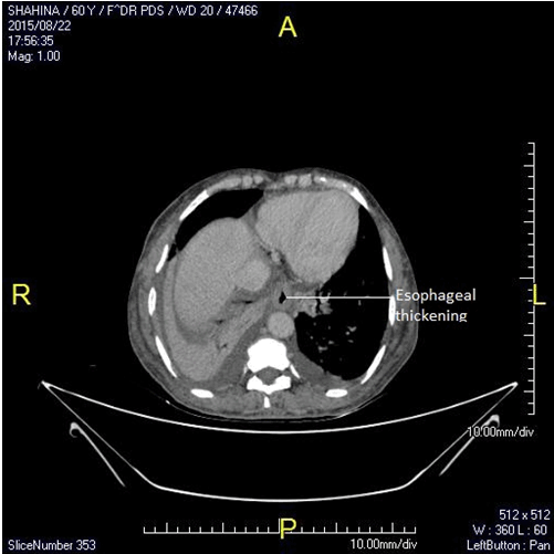
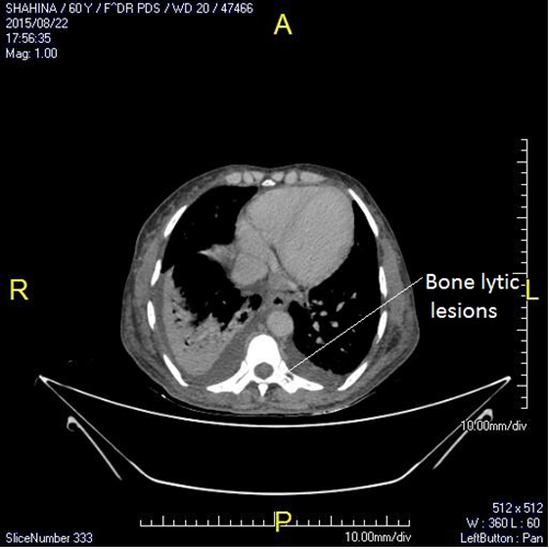
A 62-year-old female was presented to us with the chief complaint of dysphagia since six months. This was also accompanied by slurring of speech and significant weight loss of about 20 kilograms. The dysphagia was more to solids than liquids but was not associated with any regurgitation or chest pain or any manoeuvres for aiding swallowing of food. There was no history of cough, neck swelling. Patient localized the dysphagia to her throat. She was treated for leprosy one year back based on findings of a hypoaesthetic patch on the lateral side of her leg. On examination patient had pallor and macroglossia. Systemic examination was normal. On investigating she was found to have a hemoglobin of 9.2 mg/dL with an mean corpuscular volume of 72 femtolitres. Renal and liver function tests were normal. Thyroid function tests were normal. Chest X-ray showed cardiomegaly. Oesophagogastroduodenoscopy was done which showed a small hiatus hernia. Esophageal manometry was performed which showed a hypotensive lower esophageal sphincter. Computed tomography (CT) scan of chest with oral and intravenous contrast was done which showed 5 mm thickening of lower esophagus (Figure 1). Multiple lytic lesions were seen in the base of skull, spine, pelvic bones (Figure 2). Lower esophageal biopsy was normal and negative for amyloid by Hematoxylin and Eosin and Congo red staining. Based on CT findings, we evaluated the patient for leukemia, multiple myeloma or a primary carcinoma with bone metastasis. Peripheral smear examination was normal. Whole body positron emission tomography scan was negative for malignancy. Serum protein electrophoresis and urine for Bence Jones proteins were negative for myeloma. Biopsy of the bone lytic lesion showed small clusters of round cells with a plasmacytoid appearance. Immunohistochemistry was positive for multiple myeloma. We suspected the dysphagia to be secondary to amyloidosis. Patient was not willing for a tongue biopsy. Based on ultrasonography findings of enlarged kidneys and 24 hour urine protein levels of 514 mg/dl renal biopsy was performed which was positive for amyloid. Patient was started on a bortezomib/ melphalan/ prednisolone based regimen but succumbed due to cardiac failure during chemotherapy with in a few weeks. | ||||||
| ||||||
| ||||||
|
Discussion
| ||||||
|
Multiple myeloma is a neoplastic plasma-cell disorder that is characterized by clonal proliferation of malignant plasma cells in the bone marrow, monoclonal protein in the blood or urine, and associated organ dysfunction [4]. The median age at diagnosis is approximately 70 years. The diagnosis of myeloma is based on the presence of at least 10% clonal bone marrow plasma cells and monoclonal protein in serum or urine. In patients with true nonsecretory myeloma, the diagnosis is based on the presence of 30% monoclonal bone marrow plasma cells or a biopsy-proven plasmacytoma [5]. Myeloma is classified as asymptomatic or symptomatic, depending on the absence or presence of myeloma-related organ or tissue dysfunction, including hypercalcemia, renal insufficiency, anemia, and bone disease [6]. Anemia is present in about 73% of patients at diagnosis, is generally related to myeloma marrow infiltration or renal dysfunction [7]. Bony lesions develop in almost 80% of patients with newly diagnosed disease [8]. Multiple myeloma presenting as dysphagia is extremely rare and is secondary to either amyloidosis or due to an extramedullary plasmacytoma involving the pharynx or esophagus [9]. Amyloidosis can cause dysphagia due to macroglossia [1], external compression by an amyloid goitre [2] or infiltration of the lower esophageal sphincter causing pseudoachalasia [3]. Amyloid can cause infiltration of the smooth muscle of the esophagus causing pseudoachalasia which is detected on esophageal manometry by a weak peristalsis of the esophagus and poor relaxation of the lower esophageal sphincter. Endoscopic findings may be ulcerative, protusions or scirrhous. Biopsy of the esophagus may be positive for amyloid deposition in the muscularis mucosae and submucosa [10]. In view of CT findings of thickening of lower esophagus patient was subjected to lower esophageal biopsy. However, biopsy was negative for amyloid. Esophageal manometry was done which was not suggestive of achalasia. Hence we had hyothesised that the dysphagia was secondary to macroglossia. Presence of amyloid was confirmed on kidney biopsy as patients had enlarged kidneys and proteinuria. The patient was started on chemotherapy for multiple myeloma. Macroglossia can be managed with subtotal glossectomy if it is severely symptomatic causing dysphagia or stridor, albeit with a high bleeding risk [11]. However, our patient succumbed while on chemotherapy. | ||||||
|
Conclusion
| ||||||
|
Dysphagia is one of the common clinical condition. After ruling out common cause rare causes should be looked for. The whole clinical scenario should be kept in mind while evaluating any patient with dysphagia. | ||||||
|
References
| ||||||
| ||||||
|
[HTML Abstract]
[PDF Full Text]
|
|
Author Contributions
Kaivan Shah – Substantial contributions to conception and design, Acquisition of data, Analysis and interpretation of data, Drafting the article, Revising it critically for important intellectual content, Final approval of the version to be published Alisha Chaubal – Analysis and interpretation of data, Revising it critically for important intellectual content, Final approval of the version to be published Ruchir Patel – Analysis and interpretation of data, Revising it critically for important intellectual content, Final approval of the version to be published Vikas Pandey – Analysis and interpretation of data, Revising it critically for important intellectual content, Final approval of the version to be published Meghraj Ingle – Analysis and interpretation of data, Revising it critically for important intellectual content, Final approval of the version to be published Prabha Sawant – Analysis and interpretation of data, Revising it critically for important intellectual content, Final approval of the version to be published |
|
Guarantor of submission
The corresponding author is the guarantor of submission. |
|
Source of support
None |
|
Conflict of interest
Authors declare no conflict of interest. |
|
Copyright
© 2016 Kaivan Shah et al. This article is distributed under the terms of Creative Commons Attribution License which permits unrestricted use, distribution and reproduction in any medium provided the original author(s) and original publisher are properly credited. Please see the copyright policy on the journal website for more information. |
|
|