| Table of Contents | |
|
Clinical Image
| ||||||
| Acute esophageal necrosis in an end-stage renal disease patient: Endoscopic images | ||||||
| Inderpreet Grover1, Naveed Ahmad2 | ||||||
|
1MD, Department of Medicine, GV (Sonny) VAMC, Jackson, MS.
2MD, Gastroenterology, Indiana University Health Arnett, Lafayette, IN. | ||||||
| ||||||
|
[HTML Full TExt]
[PDF Full Text]
[Print This Article]
[Similar article in Pumed] [Similar article in Google Scholar] |
| How to cite this article |
| Grover I, Ahmad N. Acute esophageal necrosis in an end-stage renal disease patient: Endoscopic images. Edorium J Gastroenterol 2014;1:1–3. |
|
Case Report
| ||||||
|
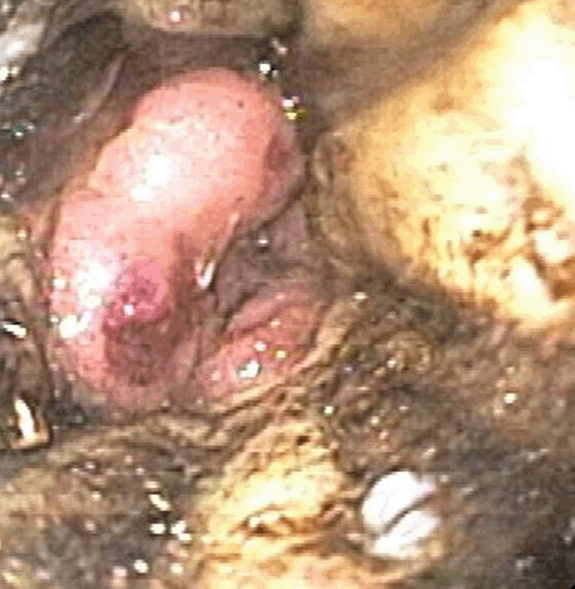
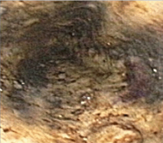
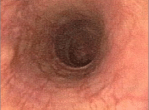
We report a case of a 43-year-old male with end-stage renal disease on hemodialysis, a history of bilateral nephrectomies, status postrenal transplant with subsequent allograft rejection, and allograft nephrectomy with multiple episodes of allograft site abscesses. He was admitted with septicemia from the abscess site, and ten days later had coffee ground emesis, requiring endoscopic evaluation. Physical examination of the patient showed pale looking with tenderness to palpation over epigastric region. The laboratory tests were significant for hemoglobin of 9 g/dL, platelets 165x103/mL, INR 1.01. Endoscopy revealed black, necrosed esophageal mucosa starting gradually at 18 cm from the incisors and progressively worsening until 40 cm, where it abruptly changed to healthy-appearing mucosa at the gastroesophageal junction (GEJ) (Figure 1) and (Figure 2). Eight days after the first endoscopy, a repeat procedure was done which showed healing of the necrosed esophageal mucosa (Figure 3). Biopsies taken at this time were consistent with necrotizing esophagitis. Figure 1 and Figure 2 show distal third of esophagus with black discoloration from acute esophageal necrosis (AEN) and normal appearing GEJ. The patient was resuscitated with fluids and was given 2 units of packed red blood cells, intravenous proton pump inhibitors were initiated resulting in complete recovery of the patient. The patient was discharged to a long-term care facility in stable condition. | ||||||
| ||||||
| ||||||
|
| ||||||
| ||||||
|
Discussion
| ||||||
|
Acute esophageal necrosis has been called by multiple names, including black esophagus and necrotizing esophagitis, owing to its acute presentation and a characteristic, circumferential, black discoloration of the esophagus, with or without exudates, that is only seen at endoscopy. Distal esophageal involvement can extend proximally, but ends at the GEJ [1]. Most common presentation of AEN is hematemesis and melena [1]. Histologic findings consistent with diffuse and severe necrosis of the mucosa and submucosa are observed in this disorder, which occurs in the absence of dysplasia, thrombosed vessels, or an inciting event, such as injury caused by acid/caustic ingestion [2]. The pathogenesis of AEN remains unknown. Some data suggest that esophageal ischemia secondary to temporary reduction in blood flow plays a major role in AEN by leading to extensive esophageal necrosis; with perfusion restored, the ischemia resolves rapidly, and the affected mucosa recovers. Most common initial presentation is upper gastrointestinal bleeding that develops rapidly after the inciting event. Diagnosis is established with upper endoscopy. Multiple comorbid conditions, including end stage renal disease, shock due to sepsis, and graft rejection may have predisposed this patient to acute esophageal necrosis. As occurred here, most treated patients recover completely. However, without treatment mortality can reach up to 32% [1]. The management of a patient with AEN requires: restoring perfusion, timely provision of intravenous proton pump inhibitors, keeping the patient NPO, and avoiding nasogastric/orogastric tubes. Antibiotics should be considered where clinically indicated as well as correction of the underlying disorder. The most common complications include gastrointestinal bleeding, requiring evaluation by endoscopy, and esophageal stricture, which can be treated by dilation. | ||||||
|
Conclusion
| ||||||
|
Gastrointestinal bleeding and esophageal stenosis are the most common complications associated with acute esophageal necrosis. Patients with hematemesis and underlying shock require prompt gastroenterology consultation. The early recognition of acute esophageal necrosis is prudent to diagnose and initiate management to reduce mortality. | ||||||
|
References
| ||||||
| ||||||
|
[HTML Full TExt]
[PDF Full Text]
|
|
Author Contributions
Inderpreet Grover – Substantial contributions to conception and design, Acquisition of data, Analysis and interpretation of data, Drafting the article, Revising it critically for important intellectual content, Final approval of the version to be published Naveed Ahmad – Analysis and interpretation of data, Revising it critically for important intellectual content, Final approval of the version to be published |
|
Guarantor of submission
The corresponding author is the guarantor of submission. |
|
Source of support
None |
|
Conflict of interest
Authors declare no conflict of interest. |
|
Copyright
© 2014 Inderpreet Grover et al. This article is distributed under the terms of Creative Commons Attribution License which permits unrestricted use, distribution and reproduction in any medium provided the original author(s) and original publisher are properly credited. Please see the copyright policy on the journal website for more information. |
|
|
|
About The Authors
| |||
| |||
| |||